
|
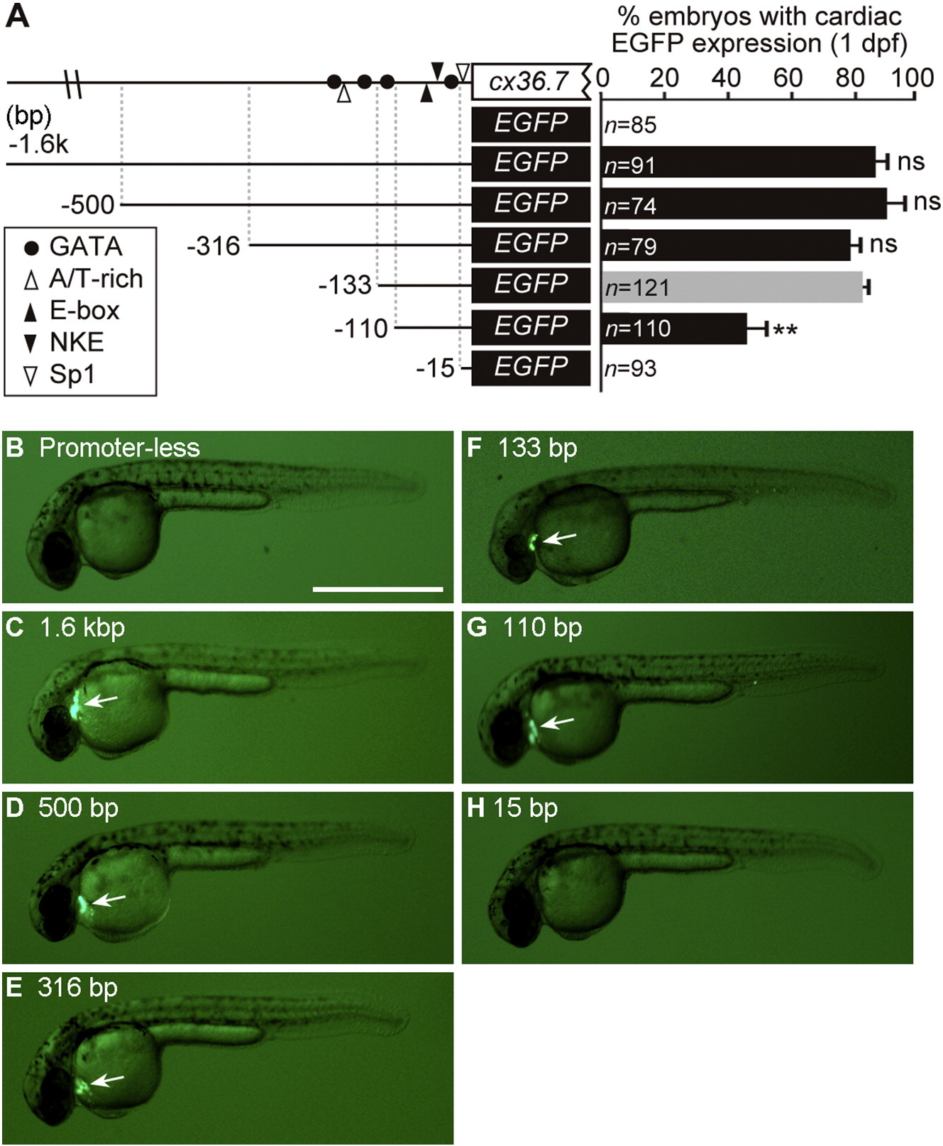
Fig. 2
Deletion analysis of the cx36.7 promoter in 1-dpf zebrafish embryos. (A) Top: schematic representation of the 5′-flanking region of the cx36.7 gene, depicting the positions of the GATA (filled circle), A/T-rich (open triangle), E-box (filed triangle), NKE (filed reversed triangle) and Sp1 (open reversed triangle) elements. Bottom: schematic representation of the 5′ deleted promoter–EGFP chimeras that were inserted between the tol2 elements in the pT2KXIGΔin vector. The reporter constructs were injected into fertilized one-cell eggs. EGFP signals in transgenic zebrafish were analyzed at 1 dpf. The bar graphs represent the percentage of embryos with cardiac-specific EGFP expression. Data are expressed as the mean ± SEM of at least three independent experiments. Significant differences for the value of the 133-bp promoter construct (**, p < 0.001) were found by one-way ANOVA with Tukey′s post-hoc test. ns, no significant difference. (B–H) Representative images of 1-dpf embryos showing no EGFP expression (B and H) or cardiac-specific EGFP expression (C–G, arrows). Bar, 1 mm.
Reprinted from Gene, 577(2), Miyagi, H., Nag, K., Sultana, N., Munakata, K., Hirose, S., Nakamura, N., Characterization of the zebrafish cx36.7 gene promoter: Its regulation of cardiac-specific expression and skeletal muscle-specific repression, 265-74, Copyright (2016) with permission from Elsevier. Full text @ Gene