
|
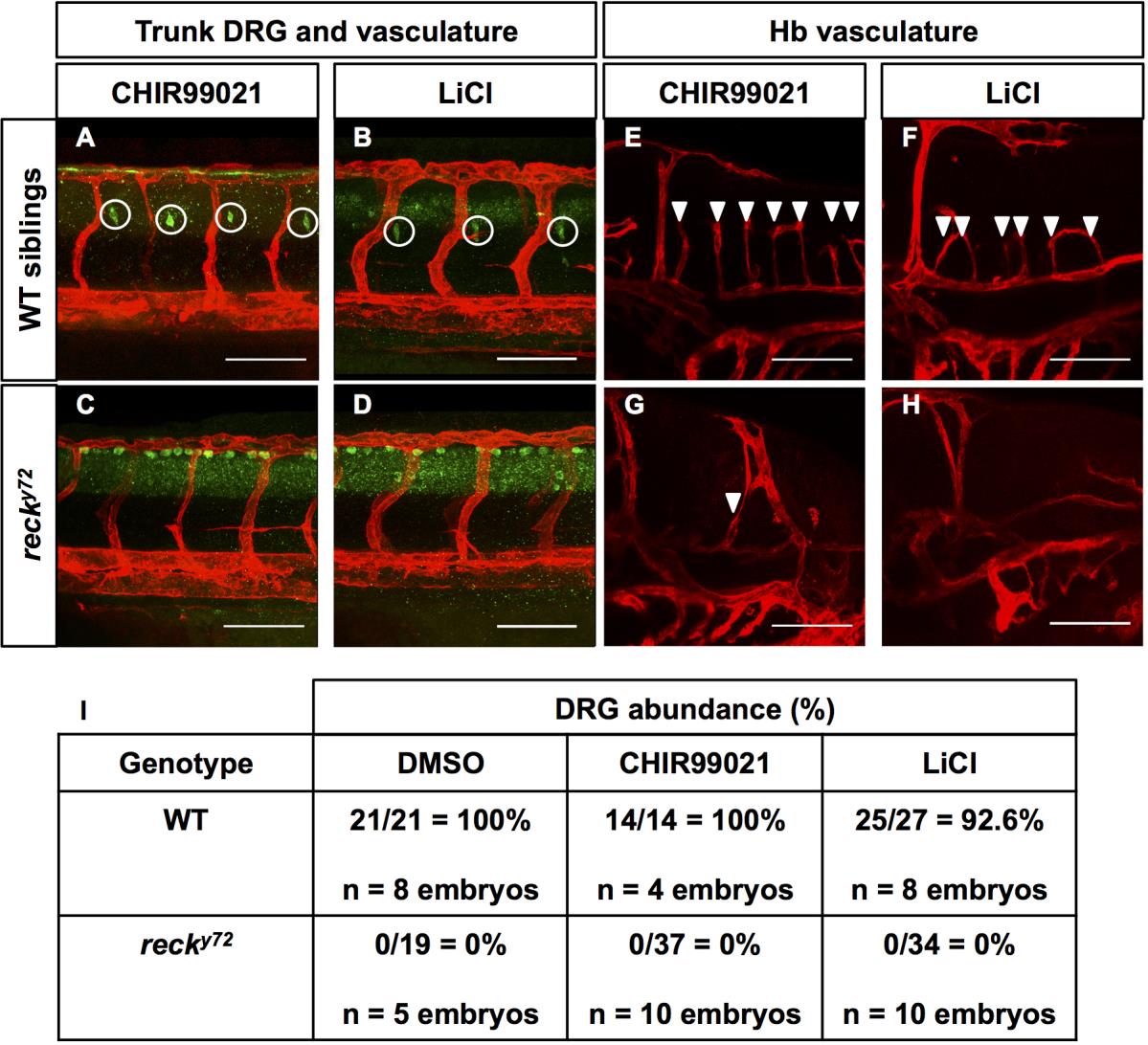
Fig. S10
Chemical activation of canonical Wnt signaling with the GSK3b inhibitors CHIR99021 and LiCl fails to rescue the DRG and CtA deficits of recky72 mutants. (A-H) Confocal lateral images at 72 hpf. Anterior, left; dorsal, up. Scale Bars: 100 µm. Genotypes and treatments as indicated. (A-D) DRG (white circles, green HuC immunofluorescence) and vasculature (Tg(kdrl:HsHRAS-mCherry)s896; red) in the trunk. (E-H) Hb vasculature (Tg(kdrl:HsHRAS-mCherry)s896; red). Central Arteries (CtA; white arrowheads). (I) Quantification of DRG abundance in WT and recky722 mutants treated with DMSO vehicle, CHIR99021 and LiCl. DRG abundance was scored by imaging a region of the anterior trunk at the level of the yolk extension spanning 3-4 Se vessels (in WT animals there is one DRG per Se vessel). Thus, the percentages were calculated from the ratio of DRG found/DRG expected.