
|
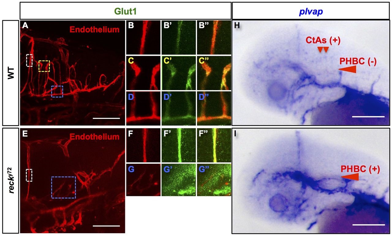
Fig. 7
reck y72 mutant embryos show aberrant cerebrovascular expression of the Wnt-responsive markers of barriergenic differentiation Glut1 and plvap. (A-G′′) Confocal lateral images of the 72hpf Hb vasculatures of WT (A-D′′) and reck y72 embryos (E-G′′). Anterior, left; dorsal, up. Endothelium, red [Tg(kdrl:RFP)s896 ]; Glut1 immunofluorescence, green. Colored dashed boxes (A,E) demarcate a region of the following vessels: white, MtA (zooms: B-B′′,F-F′′); yellow, CtAs (zooms: C-C′′); blue, PHBCs (zooms: D-D′′,G-G′′). Merged images of zooms are shown in B′′,C′′,D′′,F′′,G′′. Glut1 decorates the MtA, CtAs and PHBCs of the WT (n=7 embryos). By contrast, Glut1 decorates the MtA, but not the PHBCs, of reck y72 (n=10 embryos). (H,I) Transmitted light images of the 48hpf heads of embryos subjected to whole-mount RNA in situ hybridization with plvap riboprobes. (H) In the WT, plvap is expressed in the dorsal aspect of CtAs but not in the PHBCs (n=57 embryos). (I) In reck y72 mutants, plvap is ectopically expressed in the PHBCs (n=8 embryos). Scale bars: 100µm. Merged images of detail zooms are shown in C′′,D′′,G′′,H′′,K′′,N′′. Scale bars: 100µm. See also Figs S7-S9.