
IMAGE
Fig. S6
- ID
- ZDB-IMAGE-160224-13
- Publication
- Ulrich et al., 2016 - Reck enables cerebrovascular development by promoting canonical Wnt signaling
- All Figures
- Figures for Ulrich et al., 2016
Image
|
Figure Caption
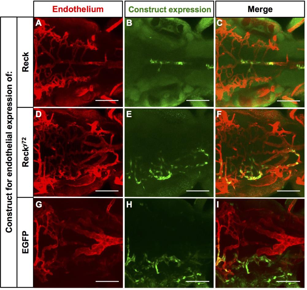
Fig. S6
Examples of the distribution of clones with exogenous endothelial expression of Reck, Recky72 or EGFP in the 72 hpf Hb vasculature of recky72 mutants. (A-I) Dorsal views (ventral-level) of the Hb extra-cerebral vasculature (red, Tg(kdrl:RFP)s896) of recky72 injected with constructs driving endothelial expression of exogenous Reck, Recky72 (both HA-tagged, see Fig. 2L) or EGFP proteins (green). Anterior, left. Right side, up. Scale Bars: 100 µm. See also Figure 5 and Graph S1.
Acknowledgments
This image is the copyrighted work of the attributed author or publisher, and
ZFIN has permission only to display this image to its users.
Additional permissions should be obtained from the applicable author or publisher of the image.
Full text @ Development