
|
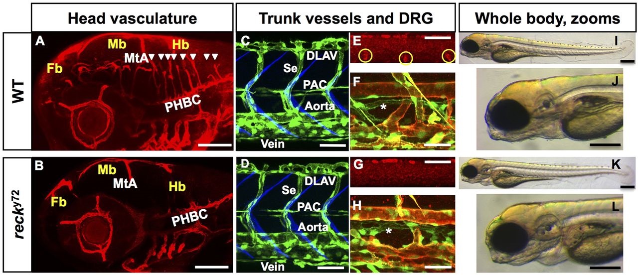
Fig. 1
nft y72 mutant embryos lack intracerebral blood vessels and DRG but have normal body morphology. Confocal (A-H) and bright-field (I-L) lateral images. Anterior, left; dorsal, up. A,B,E,G: 72 hpf; C,D: 48 hpf; F,H: 96 hpf; I-L: 60 hpf. (A,B) Central Arteries (CtAs) are found in WT (A) (white arrowheads) but are missing in nft y72 mutants (B); the other head vessels are present in nft y72. Blood vessels [Tg(kdrl:RFP)s896], red. WT (C) and nft y72 mutants (D) show identical trunk vascular patterns. Endothelium [Tg(fli1a:eGFP)y1], green; somite boundaries, blue (Zygmunt et al., 2011). DRG (yellow circles; red HuC immunofluorescence) are present in WT (E) but absent in nft y72 mutants (G). (F,H) Blood vessels are green [Tg(fli1a:eGFP)y1] and red [Tg(kdrl:RFP)s896]; lymphatics (asterisks) are green only [Tg(fli1a:eGFP)y1]. (J,L) Close-ups of head in I,K. MtA, metencephalic artery; PHBC, primordial hindbrain channel; DLAV, dorsal longitudinal anastomotic vessel; Se, intersegmental vessel; PAC, parachordal chain; Fb, forebrain; Mb, midbrain; Hb, hindbrain. Scale bars: 100µm in A,B, 50µm in C-E,G, 25µm in F,H and 200µm in I-L. See also Fig. S1, Movies 1-14 and Table S1.