
|
Fig. 1 S1
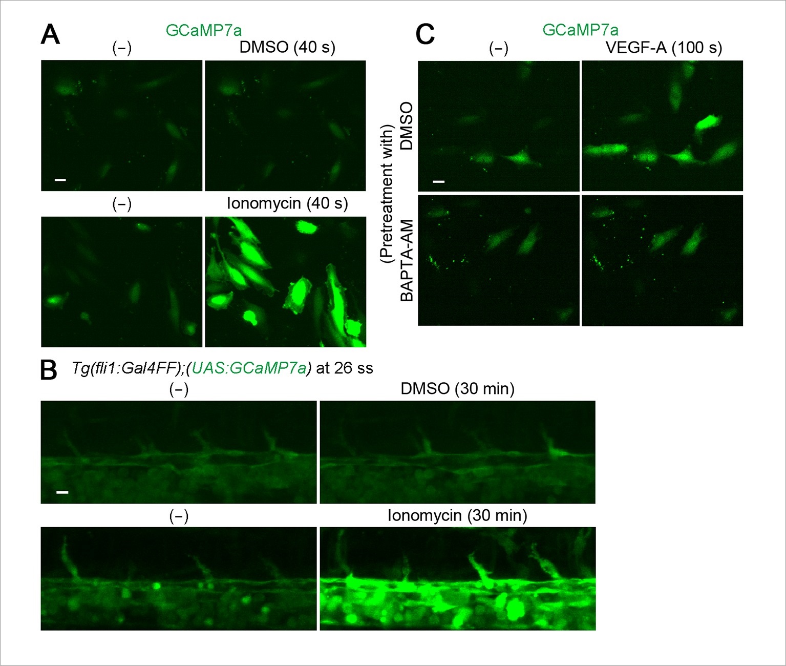
GCaMP7a works as a Ca2+ indicator in endothelial cells (ECs).
(A) HUVECs transfected with GCaMP7a expression plasmids were treated with DMSO (upper) or ionomycin (lower). GCaMP7a images before (-) and after the treatment (40 s) are shown. (B) Confocal stack fluorescence images of Tg(fli1:Gal4FF);(UAS:GCaMP7a) embryos at 26 ss treated with DMSO (upper) or 25 µM ionomycin (lower). GCaMP7a images before (-) and after the treatment (30 min) are shown. (C) Fluorescence images of HUVECs transfected with GCaMP7a expression plasmids pretreated with DMSO or 25 µM BAPTA-AM for 30 min and treated with 50 ng/ml VEGF-A before (-) and after the treatment (100 s). Note that enhancement of GCaMP7a fluorescence by VEGF-A is blocked by pretreatment with BAPTA-AM. Scale bars, 10 µm in A-C.