
|
Fig. S1
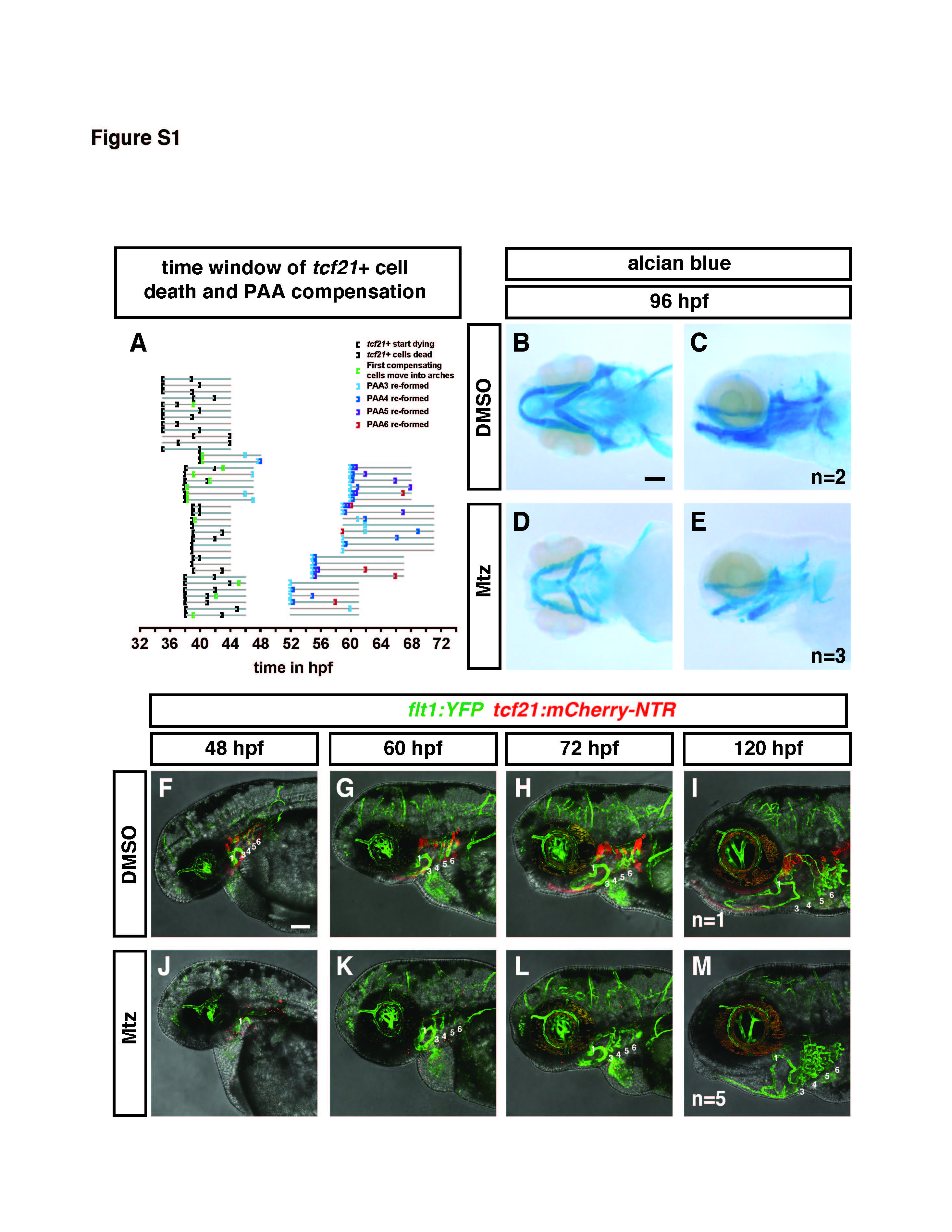
Related to Figure 3. pPAAs and cartilage form in the absence of tcf21+ cells.
(A) Graphical depiction of the time window in which tcf21+ cells die in Mtz-treated tcf21:mCherry-NTR; flk:GFP embryos and of the time window in which the PAA 3-6 recover in Mtz-treated tcf21:mCherry-NTR; flk:GFP embryos. Gray lines are timelines and indicate the length (the beginning and the end) of the movies analyzed. The open brackets indicate the beginning of dying tcf21+ cells and the beginning of the compensation (beginning of sprouting endothelial cells) in black and green, respectively. The closed brackets indicate the completion of tcf21+ cell ablation and the completion of the connection of the compensating PAA3, 4, 5 or 6 from the LDA to the VA in black and light blue, dark blue, purple and red, respectively. Please note that brackets at the beginning of a timeline of a given movie indicate that the indicated process has started or finished but that the exact time of the indicated event is before the beginning of the movie.
(B-E) Alcian blue-stained embryo at 96 hpf without (B-C) or following (D-E) treatment with Mtz. While head size is reduced in tcf21+ cell-ablated embryos, head cartilage still forms (D-E).
(F-M) Individual flt1:YFP; tcf21:mCherry-NTR embryos are followed from 48 to 120 hpf without (F-I) or following treatment with Mtz (J-M). In the control embryo, PAAs 3-6 form normally, though PAAs 3 and 4 are not labeled due to transgene variability which affects PAAs 3 and 4 in this transgenic line (F-I). In the Mtztreated embryo, PAAs 3-6 are absent at 48 hpf following ablation (J), are remade but partially lumenized at 60 and 72 hpf (K and L) and fully remade at 120 hpf (M). Number of imaged embryos is indicated. Scale bars are 100 µm.