
Fig. 5
- ID
- ZDB-IMAGE-160215-5
- Genes
- Publication
- Nagelberg et al., 2015 - Origin, specification and plasticity of the great vessels of the heart.
- All Figures
- Figures for Nagelberg et al., 2015
|
Fig. 5
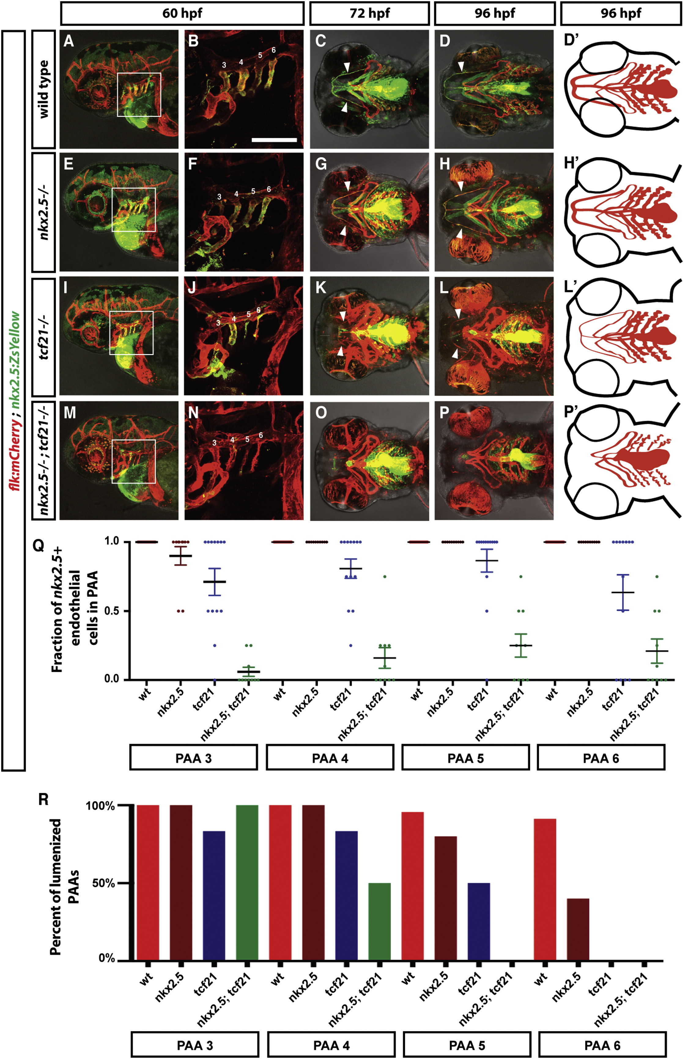
Specification of the Ventral Head Vasculature Requires the Combined Activities of Tcf21 and Nkx2.5
(A-P′) Live images and schematic diagrams of embryos of indicated stages and genotypes carrying the indicated transgenes. Squares in (A), (E), (I), and (M) indicate magnified regions shown in (B), (F), (J), and (N). Numbers indicate PAAs 3–6 in (B), (F), (J), and (N). Arrowheads indicate the HA. Lateral views (first and second column) and ventral views (third, fourth, and fifth column). Anterior is to the left. The scale bar represents 100 µm. Note that the images in (B), (F), (J), and (N) are thresholded using an ImageJ custom-written macro to only show the endothelial cells (see ImageJscripts2.ijm in Data S1).
(Q) Quantification of the contribution of nkx2.5+ cells to the PAAs 3–6 in wild-type (includes tcf21-/+; nkx2.5-/+ and tcf21-/+; nkx2.5-/+ embryos, n = 37), tcf21 mutant (includes tcf21/ nkx2.5-/+ embryos, n = 13), nkx2.5 mutant (includes nkx2.5/ tcf21-/+ embryos, n = 10), and tcf21; nkx2.5 double mutant embryos (n = 10). Individual data points and the mean with the SEM are indicated. See also Figures S2 and S3. In cases involving tcf21; nkx2.5 double mutant embryos where contribution of nkx2.5+ cells to PAAs was ambiguous due to the presence of neighboring dying nkx2.5+ cells, scoring reflects a baseline assumption of nkx2.5+ cell contribution.
(R) Quantification of lumenized PAAs of indicated genotypes at 60 hpf. The sample numbers for each genotype are: wild-type embryos (includes tcf21-/+; nkx2.5-/+ and tcf21-/+; nkx2.5-/+ embryos) n = 19, tcf21 mutants (includes tcf21/ nkx2.5-/+ embryos) n = 5, nkx2.5 mutants (includes nkx2.5/ tcf21-/+ embryos) n = 7, and tcf21; nkx2.5 double mutant embryos n = 3.