
|
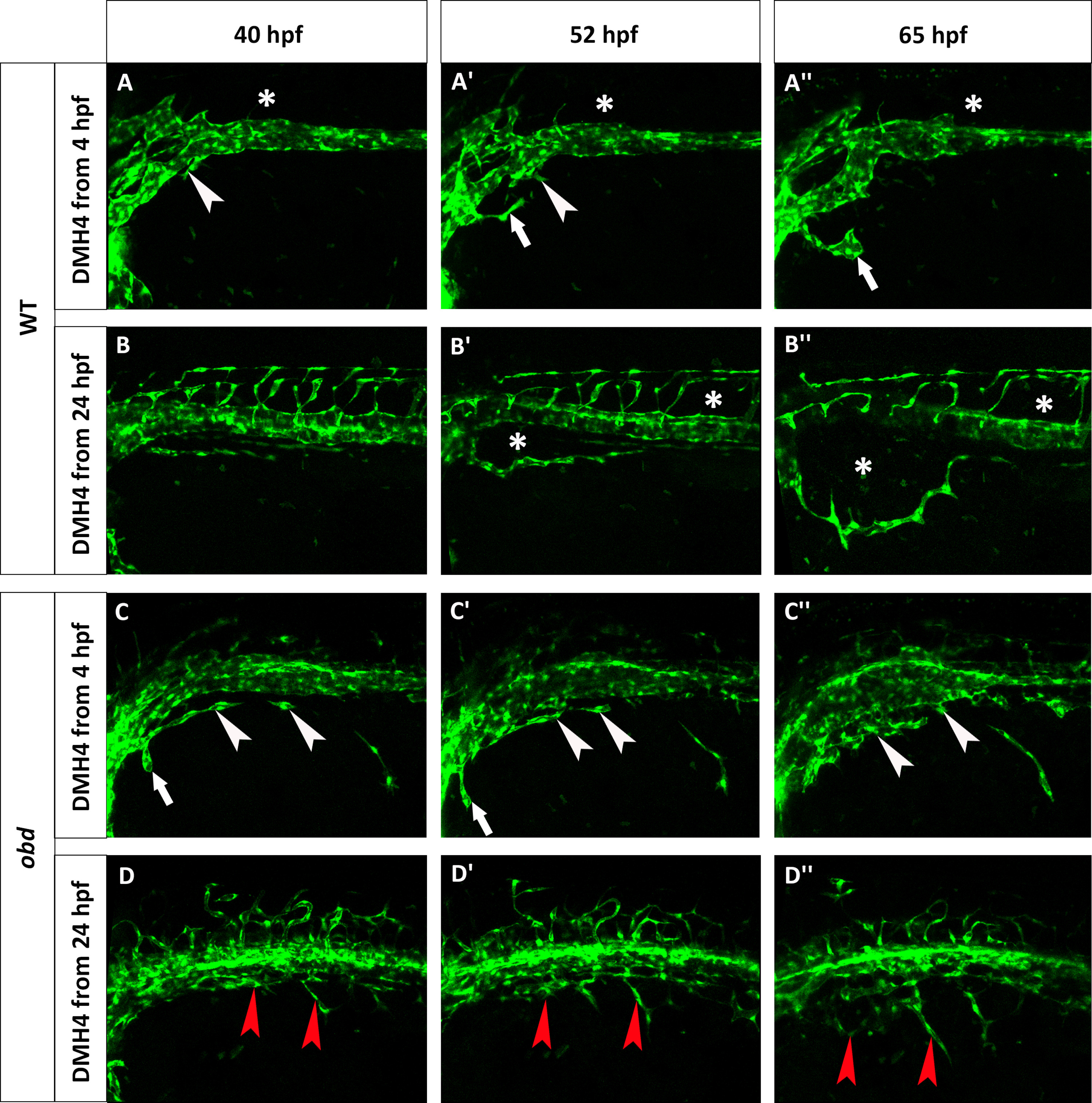
Fig. 9
obd mutants are less sensitive to Vegf inhibition. Phenotype of wild-type (A–B′′) and obd (C–D′′) embryos treated with 50 µM DMH4. (A–A′′) A Tg(fli:EGFP)y1 wild-type was treated from 4 hpf and lacks the first SIVP sprouts. White arrowheads point to the attempted sprouts from along the PCV. White arrows indicate an aberrant sprout from the duct of Cuvier. (B–B′′) A Tg(fli:EGFP)y1 wild-type was treated from 24 hpf which stops the formation of SIVP vascular compartments. Asterisks mark the absence of the internal vessels and dysmorphic ISVs. The expansion of the external vessel above the yolk ball is not affected by the inhibition. (C–C′′) obdfov01b; Tg(fli:EGFP)y1 treated embryo from 4 hpf shows some sprouts are present. White arrowheads indicate blocked sprouts from along the vein. White arrows point to irregular sprouts from the duct of Cuvier. (D–D′′) obdfov01b; Tg(fli:EGFP)y1 embryo treated from 24 hpf. Red arrowheads point to residual sprouts. Scale bars represent 100 µm.
Reprinted from Developmental Biology, 409(1), Goi, M., Childs, S.J., Patterning Mechanisms of the Sub-Intestinal Venous Plexus in Zebrafish, 114-28, Copyright (2016) with permission from Elsevier. Full text @ Dev. Biol.