
|
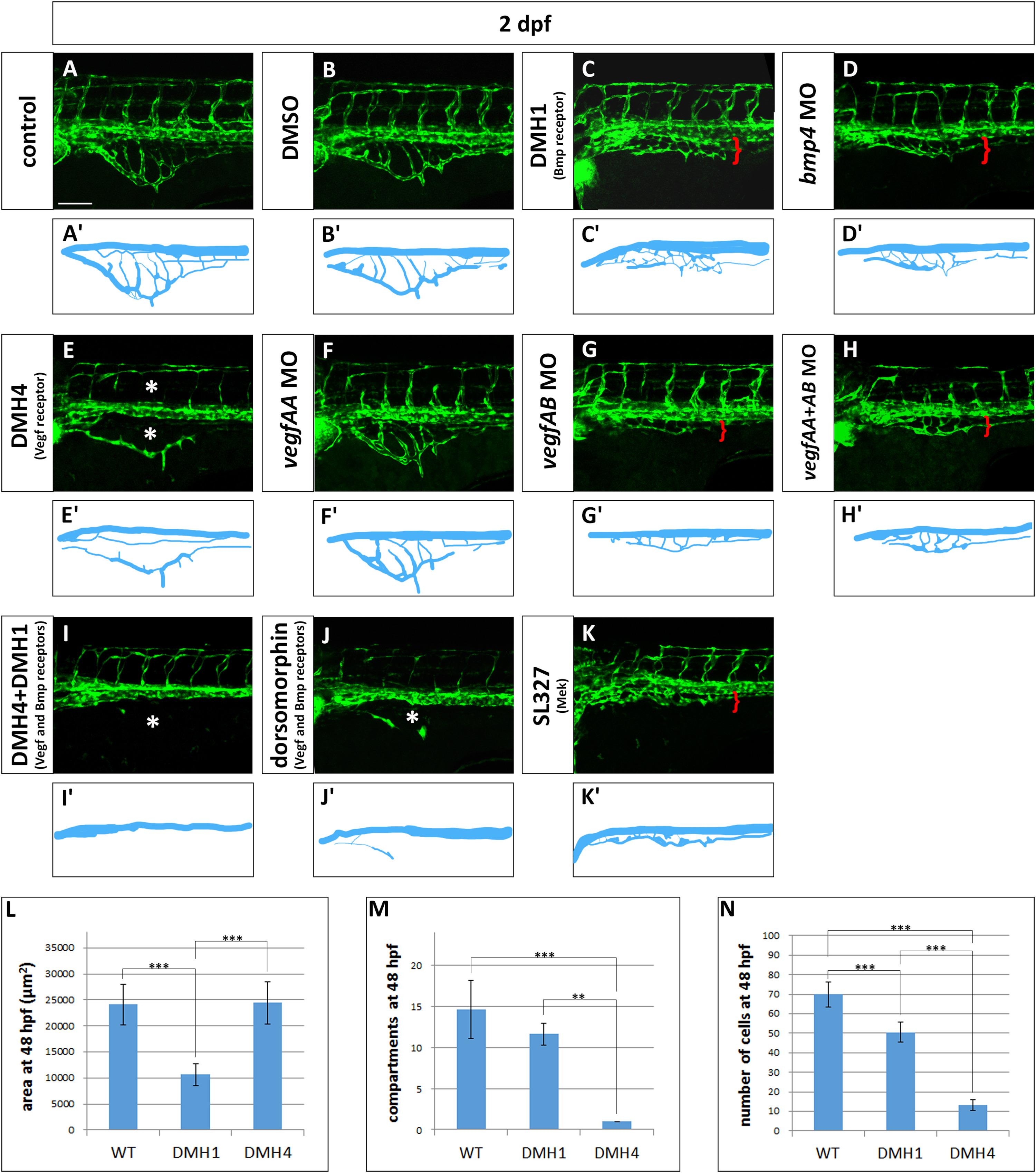
Fig. 8
Vegf and Bmp redundantly control SIVP growth. (A–K) Phenotype of embryos treated with small molecule inhibitors from 24 hpf to 48 hpf or morpholinos. (A) Untreated control embryo (B) DMSO treated control embryos. (C) 50 µM DMH1 treated embryo, an inhibitor of the Bmp type I receptor Alk2. (D) bmp4 morphant. (E) 50 µM DMH4 treated embryo, a Vegfr2 inhibitor. (F) vegfaa morphant. (G) vegfab morphant. (H) vegfaa and vegfab double morphants. (I) DMH4 and DMH1 treated embryo at 25 µM each. (J) 50 µM dorsomorphin. (K) 30 µM SL327, a Mek-1/Mek-2 inhibitor. (A′–K′) Schematics corresponding to images above (A–K). Red brackets indicate the reduced expansion of the SIVP and asterisks mark the absence of SIVP internal vessels and ISVs. Scale bar represents 100 µm. (L–N) Measurement of the average area of the vessel coverage over the yolk, number of compartments or number of cells at 48 hpf for wild-type, DMH1 treated and DMH4 treated embryos. **=p≤0.01 and ***=p≤0.001 using ANOVA.
Reprinted from Developmental Biology, 409(1), Goi, M., Childs, S.J., Patterning Mechanisms of the Sub-Intestinal Venous Plexus in Zebrafish, 114-28, Copyright (2016) with permission from Elsevier. Full text @ Dev. Biol.