
|
Fig. 7
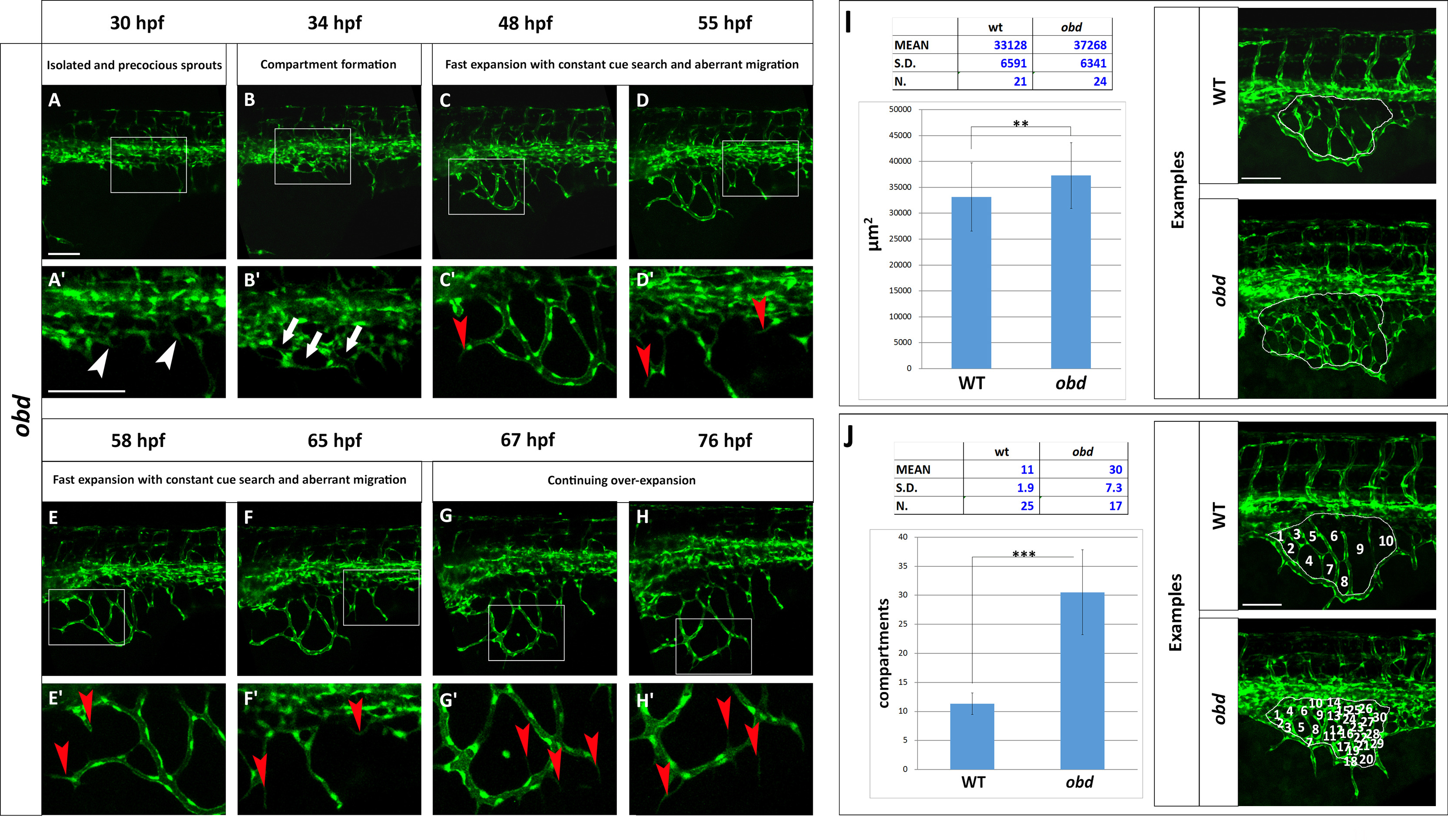
The obd SIVP shows precocious development, constant cue search, aberrant migration and higher number of compartments. (A–H) Confocal micrographs from a time-lapse of a developing obdfov01b; Tg(fli:EGFP)y1 SIVP. (A′–H′) Enlargements of images in A–H. (A′) White arrowheads indicate the precocious development of the SIVP vessels. (B2) White arrows point to some of the developing compartments. (C′–D′-E′-F′) Red arrowheads mark the presence of constant filopodial extension and aberrant migration and sprouting. (G′-H′) Red arrows indicate the formation of new filopodia scanning the environment. No pruning events are visible. (I–J) Quantitative analysis of obd SIVP vessel angiogenesis. (I) Measurement of the area of the vessel coverage over the yolk of wild-type and obd mutant embryos at 55 hpf. J) The mean number of compartments per SIVP in wild-type and obd mutant embryos was calculated at 55 hpf. The average values were calculated and plotted in a graph. **=p≤0.01 and ***=p≤ 0.001 by the Student′s T-Test. Scale bars represent 100 µm.
Reprinted from Developmental Biology, 409(1), Goi, M., Childs, S.J., Patterning Mechanisms of the Sub-Intestinal Venous Plexus in Zebrafish, 114-28, Copyright (2016) with permission from Elsevier. Full text @ Dev. Biol.