
Image
|
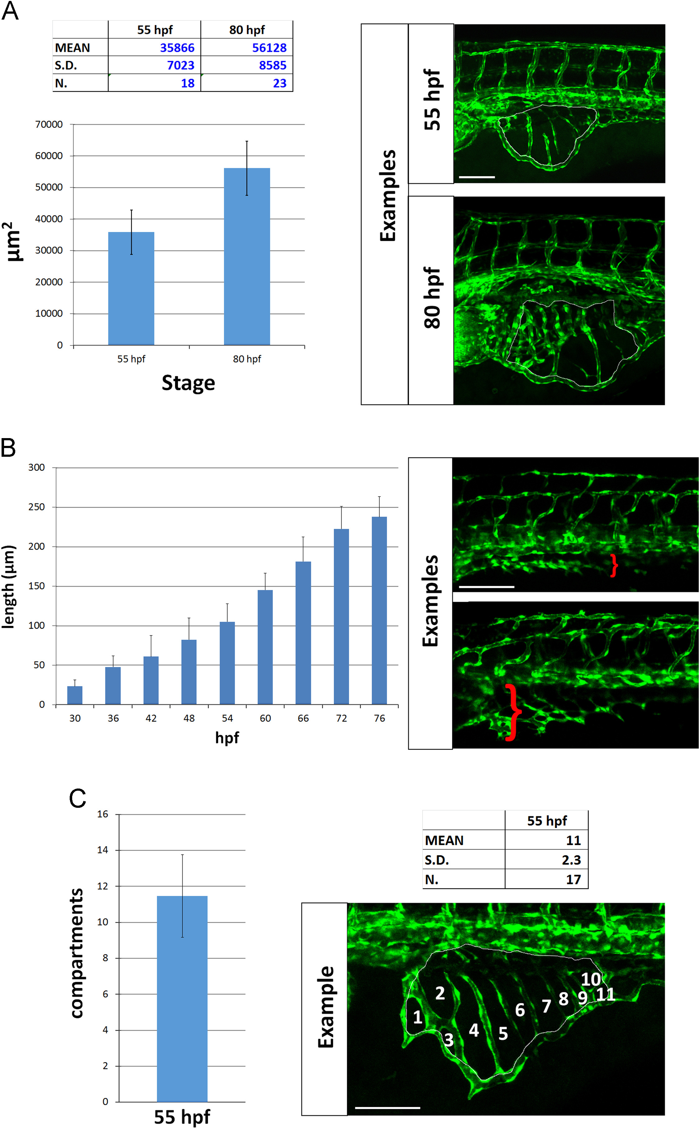
Figure Caption
Fig. 4
Quantitative analysis of SIVP vessel angiogenesis in wild-type embryos. (A) Measurement of the area of the vessel coverage over the yolk at two different stages. (B) The extent of migration of the SIVP outer basket was tracked from 30 to 76 hpf in multiple embryos. The average values were calculated and plotted in a graph. (C) The mean number of compartments per SIVP was calculated at 55 hpf. Scale bars represent 100 µm.
Acknowledgments
This image is the copyrighted work of the attributed author or publisher, and
ZFIN has permission only to display this image to its users.
Additional permissions should be obtained from the applicable author or publisher of the image.
Reprinted from Developmental Biology, 409(1), Goi, M., Childs, S.J., Patterning Mechanisms of the Sub-Intestinal Venous Plexus in Zebrafish, 114-28, Copyright (2016) with permission from Elsevier. Full text @ Dev. Biol.