
|
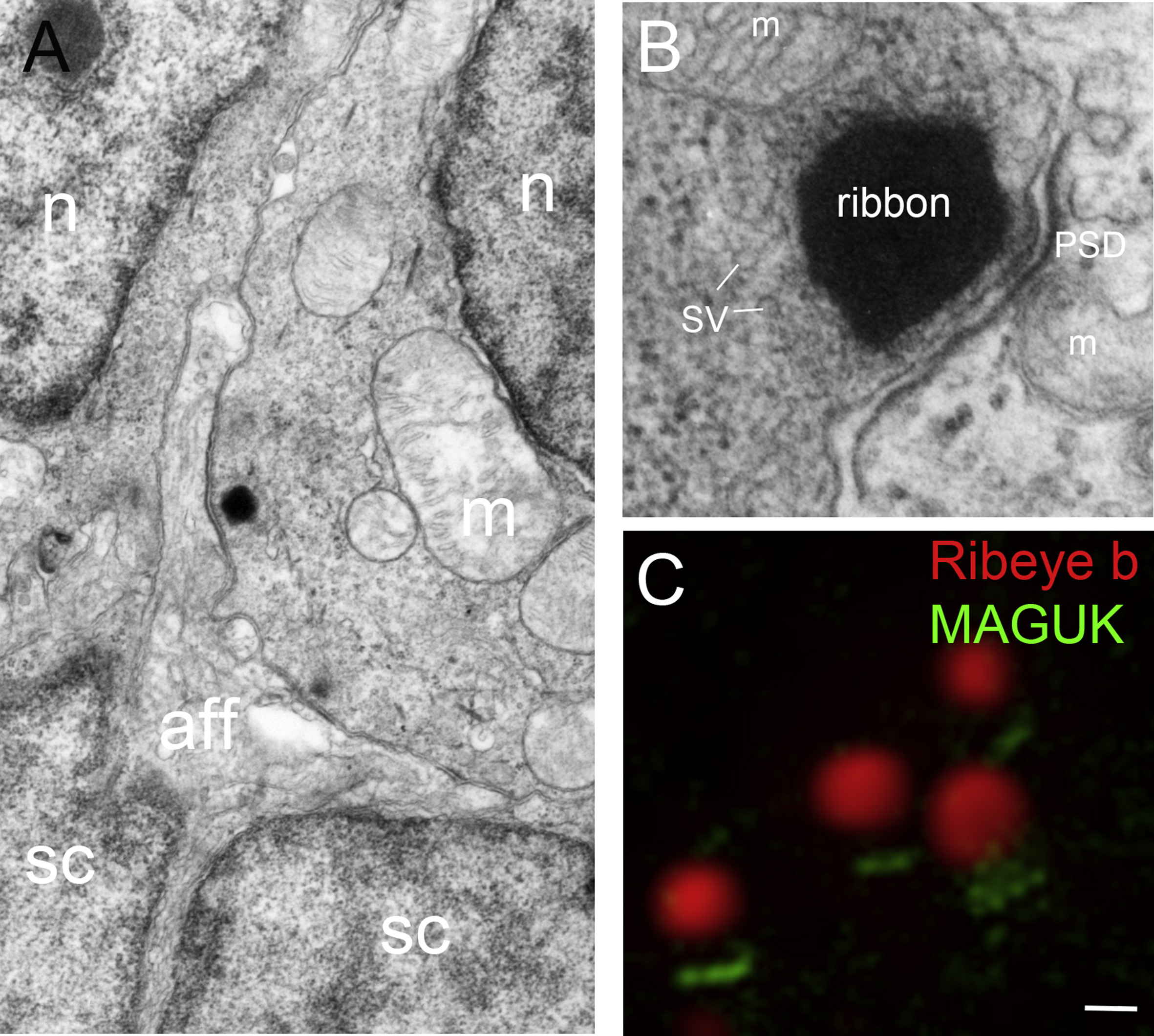
Fig. 1 Ribbon synapses in zebrafish hair cells. A, Transmission electron microscopy (TEM) micrograph of the basal end of an inner ear hair cell located within the anterior macula. A prominent ribbon with vesicles is seen juxtaposed to an afferent fiber that appears to contact a large surface area (a second faint ribbon outside of the sectioning plane is seen to right). B, High magnification view of a ribbon along with the postsynaptic density. C, Super resolution structured illumination microscopy (SR-SIM) image of several ribbon synapses labeled with Ribeye b (red) and pan-MAGUK (green) antibodies (Image: Lavinia Sheets). Abbreviations: aff, afferent; m, mitochondria; n, nucleus; PSD, postsynaptic density; sc, supporting cell; SV, synaptic vesicle. Scale bar, 200 nm in A; 75 nm in B; C, 250 nm.
Reprinted from Hearing Research, 330(Pt B), Nicolson, T., Ribbon Synapses in Zebrafish Hair Cells, 170-7, Copyright (2015) with permission from Elsevier. Full text @ Hear. Res.