
|
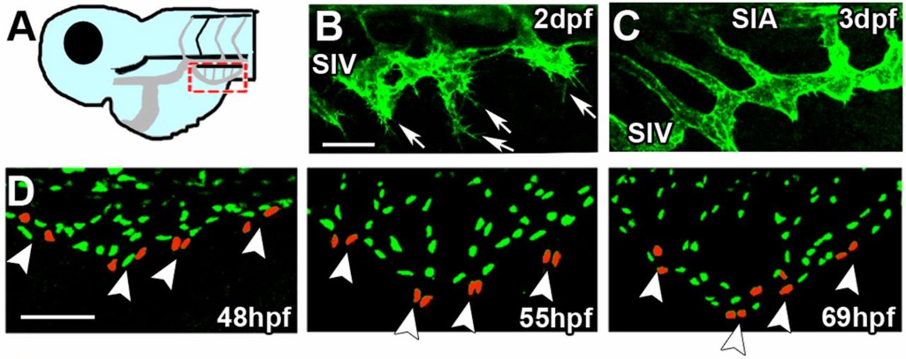
Fig. 6
Establishment of the SIV involves leading bud-guided collective migration of ECs. (A) Dashed red box in the diagram shows approximate location of regions imaged in B-D. (B) Actin-rich filopodia (arrows) are detected at the SIV migration front in Tg(Lifeact:GFP) embryos at 2dpf. (C) As the plexus reaches its stereotypical basket shape at 3dpf, all filopodia retract. (D) Distribution of EC nuclei in Tg(fli1:nGFP) embryos demonstrates that leading buds consist of paired ECs (48-55hpf, arrowheads), rather than of a single tip cell. Towards the end of the process, leading buds retract and are incorporated into the SIV (69hpf, arrowheads). Scale bars: 50µm.