
|
Fig. 5
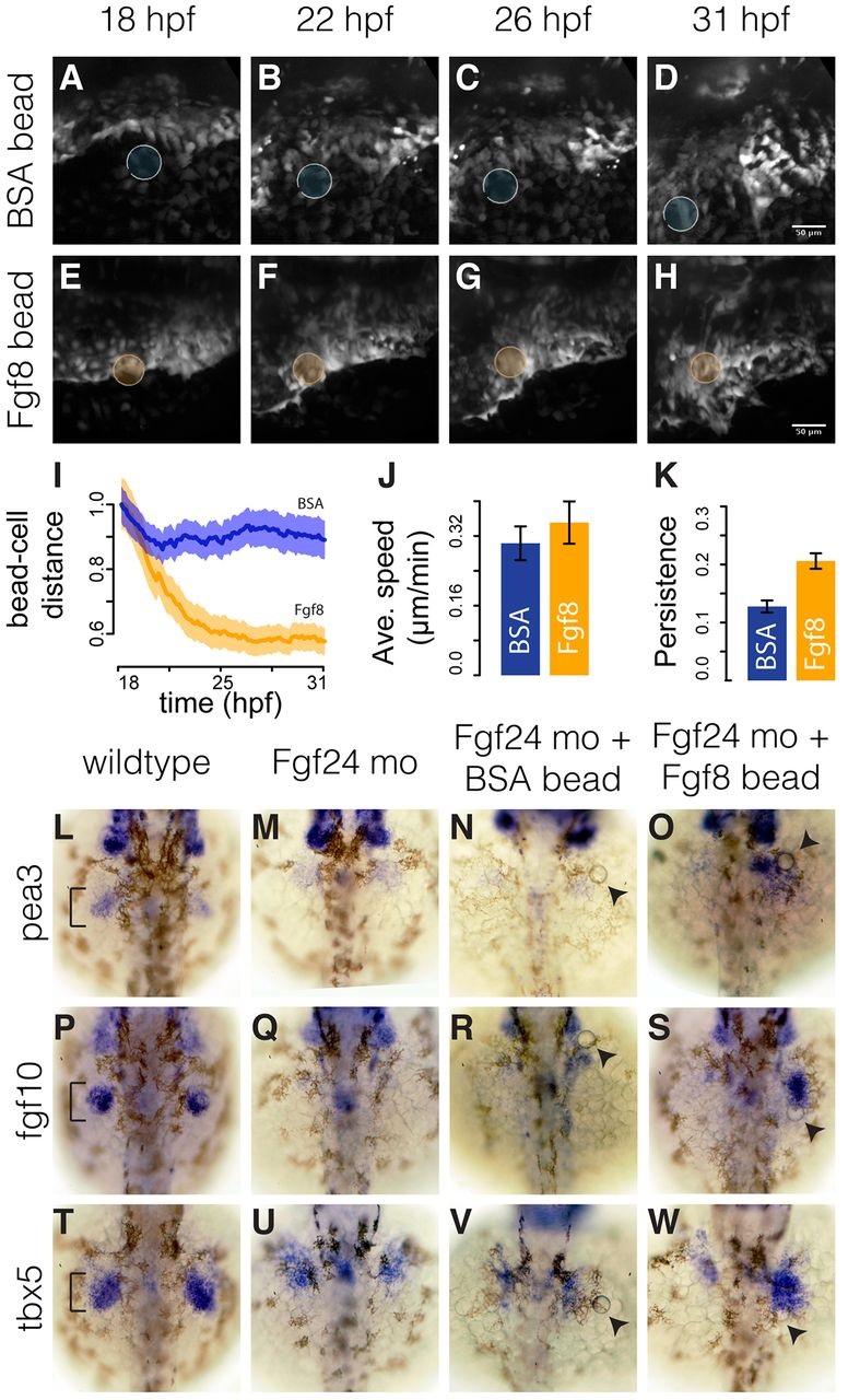
Fin-field LPM cells in Fgf24 morphants can converge towards Fgf8b-coated beads. (A-H) Stills from time-lapse of Fgf24 morphant embryos implanted with either a BSA-coated bead (A-D) or an Fgf8b-coated bead (E-H). Scale bars: 50 µm. (I) Temporal progression of relative distances of tracked cells to implanted BSA- or Fgf8b-coated beads. Solid lines show eight-embryo average. Shaded areas show eight-embryo 95% confidence interval. n=20 tracks per embryo. (J,K) Average speed (J) and persistence (K) of tracked cells in Fgf24 morphant embryos with BSA- or Fgf8b-coated beads. Error bars show eight-embryo 95% confidence interval. (L-W) Dorsal views of fin bud region (brackets) in 36hpf embryos showing pea3 (L-O), fgf10 (P-S) and tbx5a (T-W) expression in wild types, Fgf24 morphants, Fgf24 morphants with a BSA-coated bead and Fgf24 morphants with an Fgf8b-coated bead. Arrowheads point to implanted beads.