
Image
|
Figure Caption
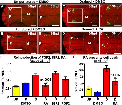
Fig. 4
RA prevents neuroepithelial cell death at 36 and 48 hpf. (A–D) Dorsal view of TUNEL (green) and propidum iodide (red) of un-punctured (A), punctured (B) or drained embryos (C) injected with DMSO and drained embryos injected with RA (D). (E–F) Quantification of cell death at 36 hpf after introduction of FGF2, IGF2, or RA (E) or at 48 hpf after introduction of RA (F). Data represented as mean ± SEM. F = forebrain, M = midbrain, UP = unpunctured, P = punctured, D = drained. Scale bars = 50 µm.
Acknowledgments
This image is the copyrighted work of the attributed author or publisher, and
ZFIN has permission only to display this image to its users.
Additional permissions should be obtained from the applicable author or publisher of the image.
Full text @ Dev. Neurobiol.