
|
Fig. 1 S3
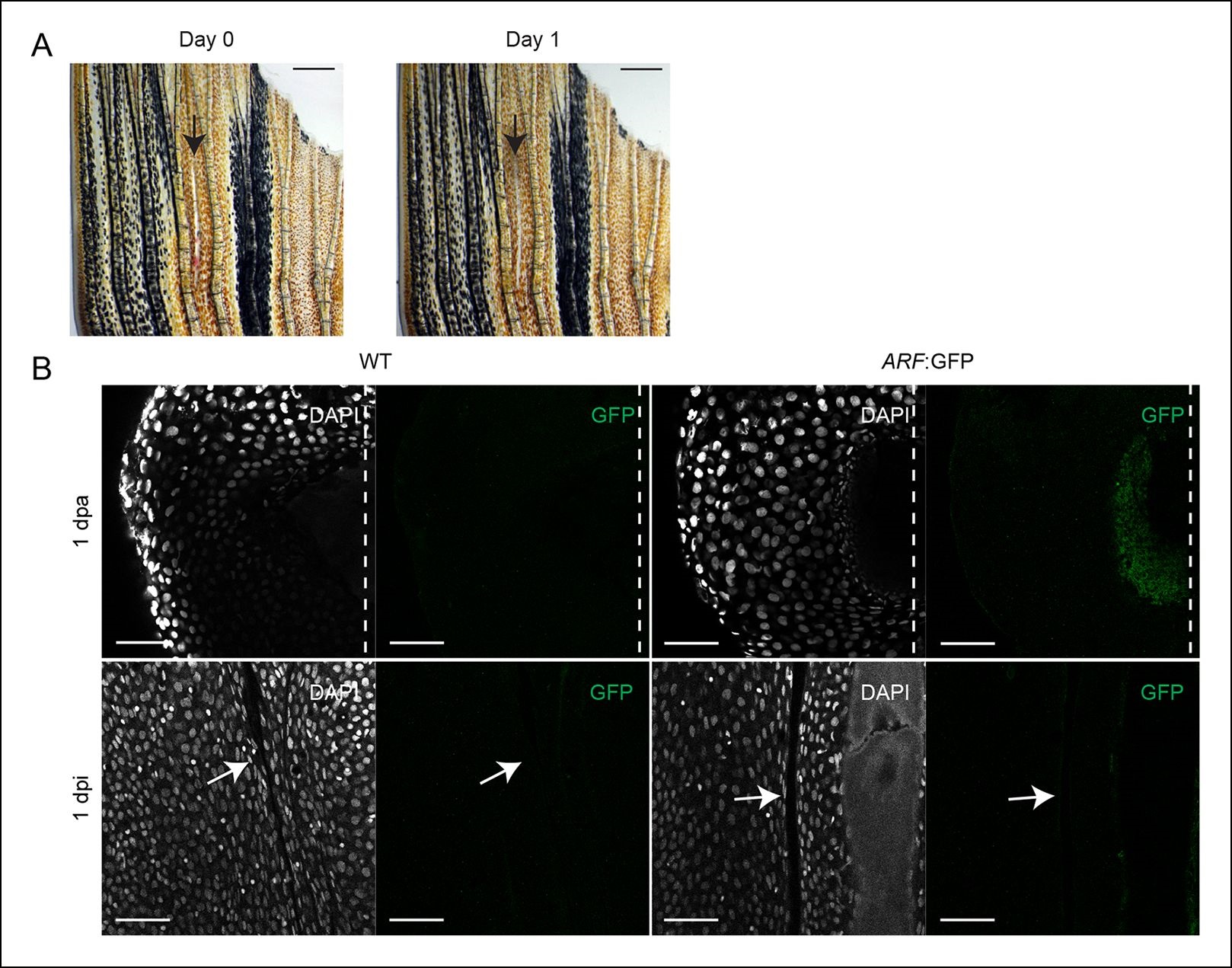
ARF is not activated during wound healing in the absence of a blastema. (A) At day 0, dorsal fin lobes were wounded (interray laceration, 0 dpi), while ventral fin lobes were amputated (0 dpa). At day 1, GFP expression was assayed in the healing (dorsal) and regenerating (ventral) fins. Scale bars: 0.5 mm. (B) Representative images (sagittal confocal images) of cytoplasmic GFP expression in WT and ARF:GFP fins that were either amputated (1 dpa) or wounded (1 dpi). GFP is only detected in ARF:GFP fins that have been amputated (N = 5). Scale bars: 50 µm. Arrows point to the interray wound. Dashed lines represent amputation planes.GFP: Green fluorescent protein; WT: Wild type.