
Fig. 4
- ID
- ZDB-IMAGE-160115-9
- Publication
- Sidik et al., 2015 - A zinc finger protein that regulates oligodendrocyte specification, migration, and myelination in zebrafish
- All Figures
- Figures for Sidik et al., 2015
|
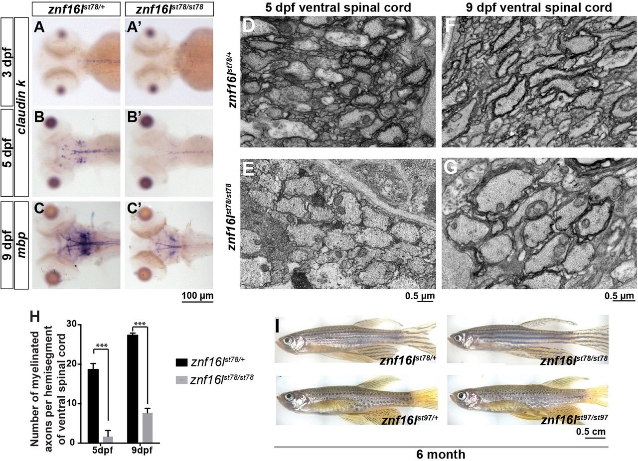
Fig. 4
CNS myelin is reduced in znf16l mutants. (A-C) Expression of mature oligodendrocyte markers claudin k (A,B) and mbp (C) was reduced in znf16lst78 mutants between 3 and 9dpf. Expression of cldnk was reduced but detectable at 5dpf (B), and expression of mbp was reduced but detectable at 9dpf (C). (D,E) Transverse transmission electron microscopy images of ventral spinal cord showed that many wild-type axons were already myelinated in wild type (D) but not mutants (E). (F,G) At 9dpf, more axons were myelinated in the mutants (G) than at 5dpf, but the number was still much less than in wild-type siblings at 9dpf (F). (H) Quantification of the number of myelinated axons in ventral spinal cord at 5 and 9dpf. Error bars show s.d.; significance was determined with two-tailed Student′s t-test. ***P<0.001 in H. (I) Adult znf16l homozygous mutants are viable and fertile, with no gross morphological defects compared with their wild-type siblings for both of the alleles. Genotypes of all fish analyzed were determined by PCR assay.