
|
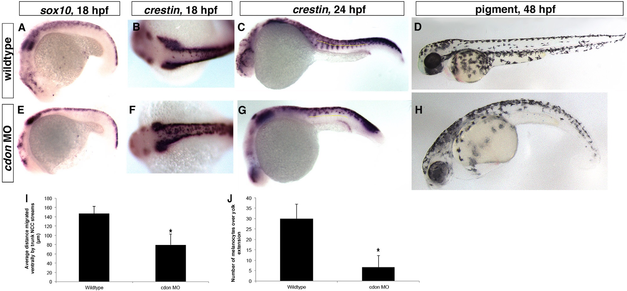
Fig. 2
cdon is required for trunk neural crest migration. Lateral views, anterior to the left except (B) and (F) which is a dorsal view with anterior to the left. (A)–(C), (E)–(G) ISH for neural crest markers sox10 at 18 somites and crestin at 18 and 24 hpf in wildtype and cdon morphant embryos. (A)–(C) In wildtype embryos, NCCs migrate in streams ventrally from the neural tube, while (E)–(G) cdon morphant NCCs do not migrate far ventrally and appear to stall after migrating a short distance (distance migrated at 24 hpf by the most ventral NCC in each of the first 7 migratory streams over the yolk extension; wildtype average=146.9 µm, cdon MO average=79.7 µm; p=0.000009). In addition more NCCs are localized along the dorsal midline, suggesting a migratory defect (compared B to F; sox10: wildtype, n=40/41 normal migration where NCC streams migrate ventral to midline in at least 5 out of the first 7 streams above the yolk extension(y.e.); cdon MO, n=33/39 with impaired migration; crestin: wildtype n=62/64 normal migration, cdon MO, n=21/25). (D) and (H) Imaging of melanocytes at 48 hpf displays a loss of ventral melanocytes in cdon morphants (average of 6.7 over yolk extension) compared to wildtype (average 30 over y.e., p=0.012), suggesting NCCs do not reach ventral positions. (I) and (J) Quantification of C, G and D, H.
Reprinted from Developmental Biology, 407(2), Powell, D.R., Williams, J.S., Hernandez-Lagunas, L., Salcedo, E., O'Brien, J.H., Bruk Artinger, K., Cdon promotes neural crest migration by regulating N-cadherin localization, 289-99, Copyright (2015) with permission from Elsevier. Full text @ Dev. Biol.