
|
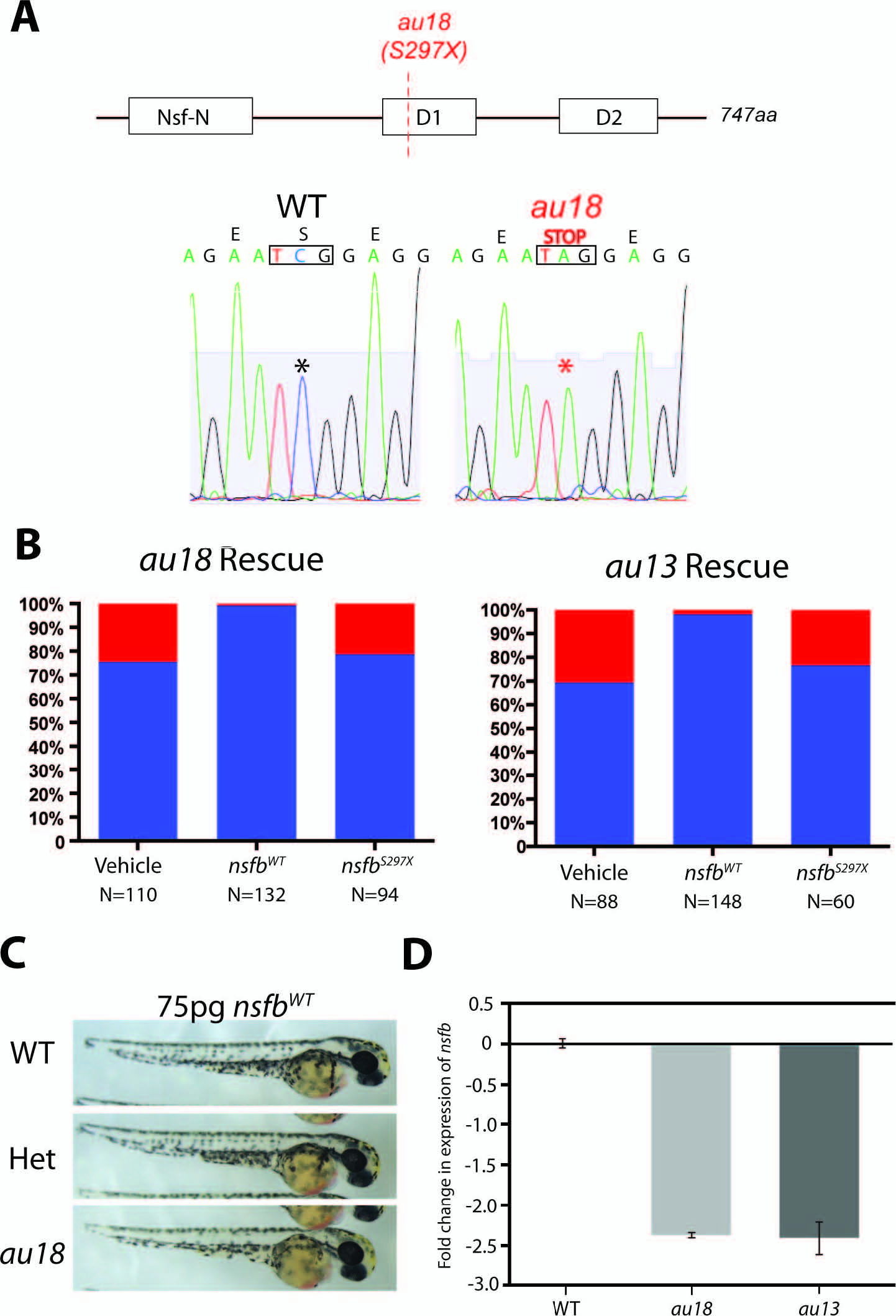
Fig. 3 au13 and au18 phenotypes are likely caused by mutations in nsfb. (A) Schematic of the nsfb protein. There are three major domains: the Nsf-N, ATPase domain 1 (D1), and ATPase domain 2 (D2). The mutation occurs at nucleotide 893 (C893T), and the dotted red line indicates the location of the premature stop codon at amino acid 297 (S297X) in au18 mutants. This mutation is predicted to truncate the protein by 60%. (B) Graph illustrating the percentage of phenotypically mutant embryos in each clutch following injection of vehicle, nsfbWT, and nsfbau18 mRNA. Both au13 and au18 mutant phenotypes are rescued by injection of nsfbWT, whereas injection of nsfbau18 mRNA failed to rescue either the au13 or au18 mutant phenotype. (C) Representative images of WT, au18+/-, and au18-/- embryos following nsfbWT injection. (D) Quantitative PCR quantification of the relative fold change of nsfb in nsfbau13 and nsfbau18 embryos at 48 hpf. Transcript levels were normalized to act2b. Error bars indicate SEM of five technical replicates for each experiment. n = 3 biological replicates.