
Image
|
Figure Caption
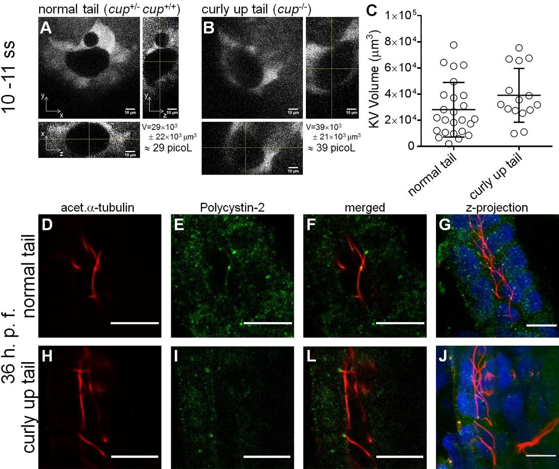
Fig. S1 cup mutant KV volumes. Whole KV live-microscopy scans of 10–11 s.s. embryos from cup+/- ; foxj1a:GFP parents. The middle focal plan along the XY axis and the respective orthogonal views (along XZ and YZ axes) are shown for the most representative normal tail (cup+/-; cup+/+) (A), and curly-up tail (cup-/-) (B) embryos. KVvolume is indicated in µm3 and in picoL. Scale bars: 10 µm. (C) Estimated KV volumes (µm3) for normal tail (cup+/-; cup+/+) (n = 23) and curly-up tail (cup-/-) (n = 15) embryos. Average values and the respective s.d. are indicated.
Figure Data
Acknowledgments
This image is the copyrighted work of the attributed author or publisher, and
ZFIN has permission only to display this image to its users.
Additional permissions should be obtained from the applicable author or publisher of the image.
Full text @ Biol. Open