
|
Fig. 2
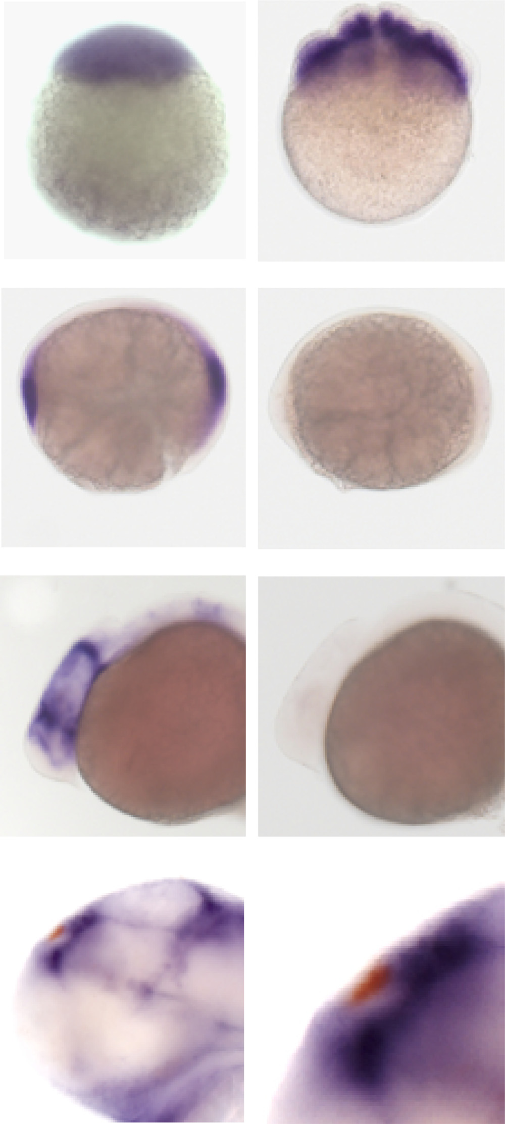
wls is maternally deposited and not expressed in the dorsal habenular nuclei. Maternally derived wls transcripts are detected at the (A) 1-cell and (B) 8-cell stage. All embryos derived from heterozygous (wlsc186/+) parents have wls transcripts; however, by (C) 90% epiboly, they are no longer detected in 25% of the progeny. (D) Expression of wls in the diencephalon and midbrain–hindbrain boundary at 24 hpf is not observed in homozygous wls mutants. (E) wls transcripts (blue) are found in cells surrounding but not within the dorsal habenular nuclei (red, indicated by arrowhead in boxed area shown on right). (For interpretation of the references to color in this figure legend, the reader is referred to the web version of this article.)
Reprinted from Developmental Biology, 406(2), Kuan, Y.S., Roberson, S., Akitake, C.M., Fortuno, L., Gamse, J., Moens, C., Halpern, M.E., Distinct requirements for Wntless in habenular development, 117-28, Copyright (2015) with permission from Elsevier. Full text @ Dev. Biol.