
|
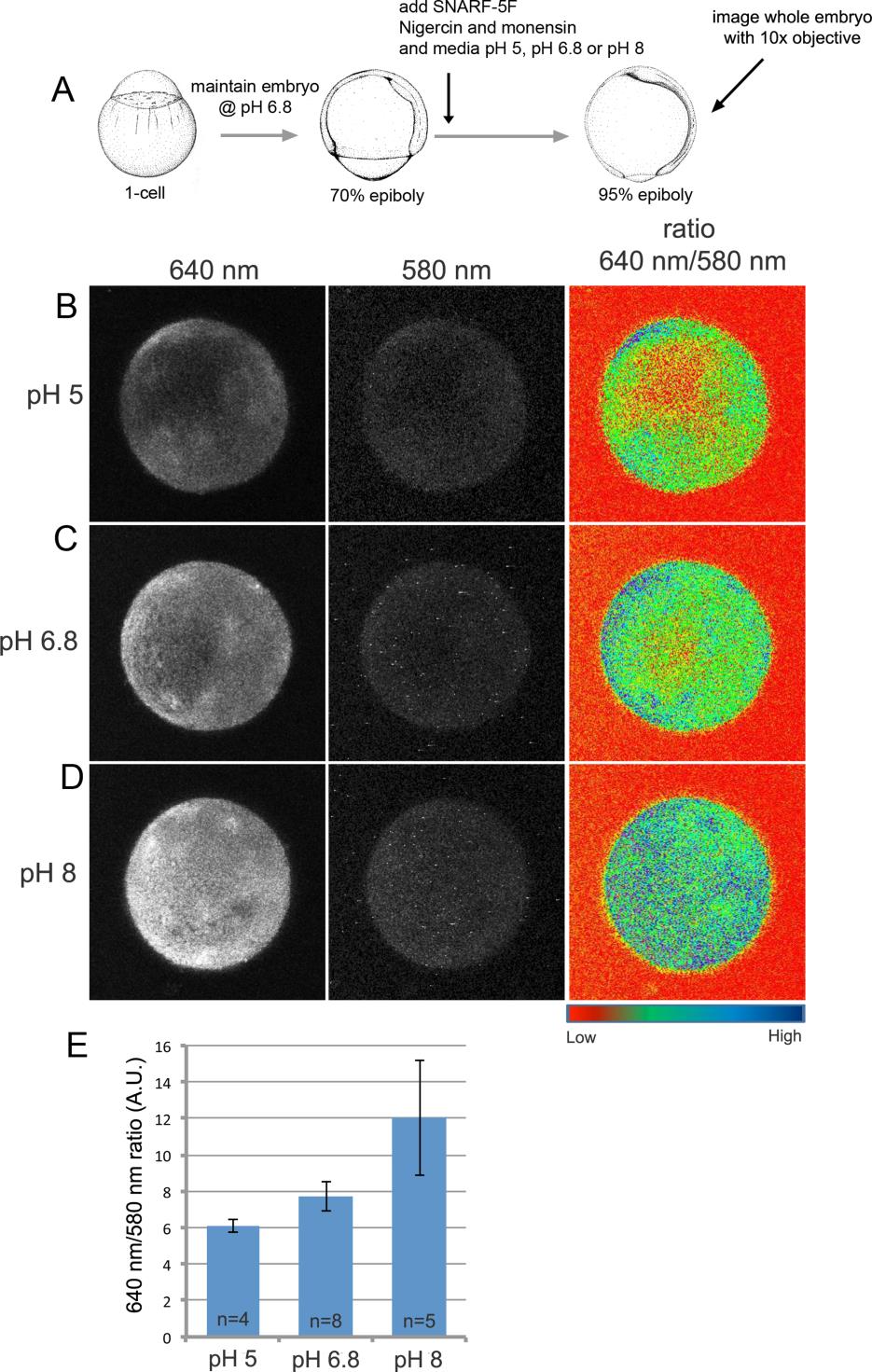
Fig. S6 SNARF-5F displays pH-dependent fluorescence in the zebrafish embryo. (A) Cartoon of experimental design to validate SNARF-5F utility in zebrafish. (B-D) Fluorescent images of entire wild-type embryos treated with nigercin and monensin and then maintained at pH 5 (B), pH 6.8 (C) or pH 8 (D). Fluorescence emission at 640 nm increased with pH, whereas emission at 580 nm was pH-independent and served as a dye loading control. A heat map of the 640 nm to 580 nm ratio revealed pH-dependent intensity differences. (E) Average 640 nm to 580 nm ratios show a consistent pH-dependent increase of SNARF-5F fluorescence. A. U.=arbitrary units.
Reprinted from Developmental Biology, 407(1), Gokey, J.J., Dasgupta, A., Amack, J.D., The V-ATPase accessory protein Atp6ap1b mediates dorsal forerunner cell proliferation and left-right asymmetry in zebrafish, 115-30, Copyright (2015) with permission from Elsevier. Full text @ Dev. Biol.