
|
Fig. 7
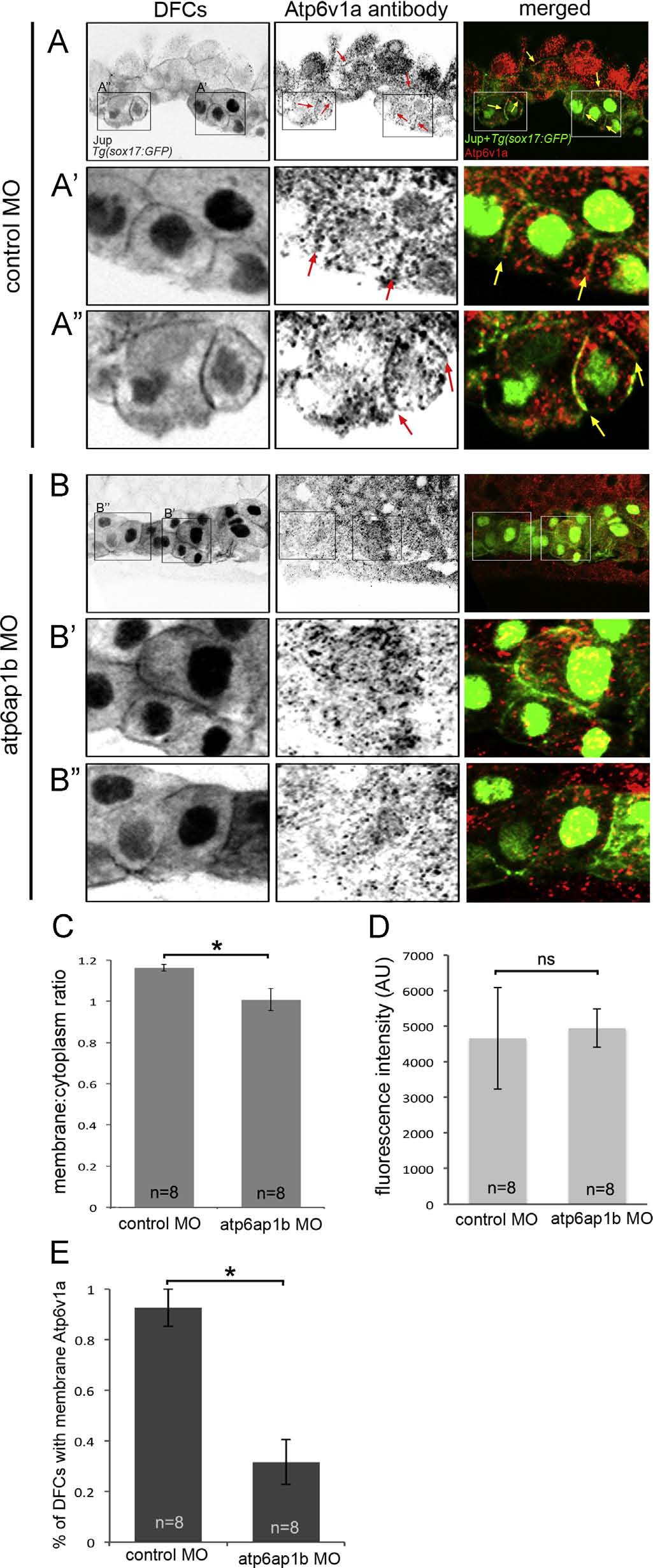
Atp6v1a localization in DFCs is altered in Atp6ap1b depleted embryos. (A,B) Confocal sections through a subset of DFCs labeled with Atp6v1a antibodies. Punctate Atp6v1a signals were detected in the cytoplasm and along some plasma membranes (arrows) marked by Jup antibodies in DFCs in Tg(sox17:GFP) embryos injected with control MO (A). A′ and A′′ show enlarged images of the boxed regions in A. Plasma membrane association of Atpv1a signals was reduced in Atp6ap1b MO embryos (B). B′ and B′′ show enlarged images of the boxed regions in B. (C) A plasma membrane-to-cytoplasm ratio of Atp6v1a fluorescence in DFCs. (D) Overall Atpv1a fluorescence in DFCs. (E) The percentage of DFCs found to have Atp6v1a puncta associated with Jup staining at the plasma membrane.
Reprinted from Developmental Biology, 407(1), Gokey, J.J., Dasgupta, A., Amack, J.D., The V-ATPase accessory protein Atp6ap1b mediates dorsal forerunner cell proliferation and left-right asymmetry in zebrafish, 115-30, Copyright (2015) with permission from Elsevier. Full text @ Dev. Biol.