
|
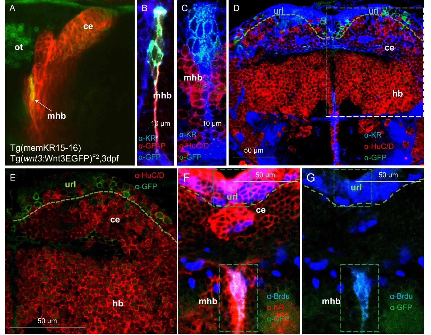
Fig. S2 Cells expressing Wnt3EGFP in the midbrain hindbrain boundary and cerebellum are located in proliferation niches. (A) An in vivo image of a double transgenic larvae co-expressing the membrane localized KillerRed in the midbrain hindbrain boundary (mhb) and cerebellum of Tg(memKR15-16) and C terminal EGFP Wnt3 fusion proteins from Tg(wnt3:Wnt3EGFP)F2. Double transgenic larvae confirmed Wnt3EGFP expression in the mhb and the cerebellum. wnt3:Wnt3EGFP+ cells in the cerebellum are predominantly localized to the upper rhombic lip (url). (B-E)Wnt3EGFP cells at midbrain hindbrain boundary (mhb) are glial cells (B) as it stained positively for GFAP (glial fibrilary acidic protein) and are negative for the neuronal marker HuC/D (C-E). (F-G) Midline restricted Wnt3EGFP cells at midbrain hindbrain boundary (demarcated by membrane KR expression in this domain) are proliferating cells since they stained positively for Brdu.