
|
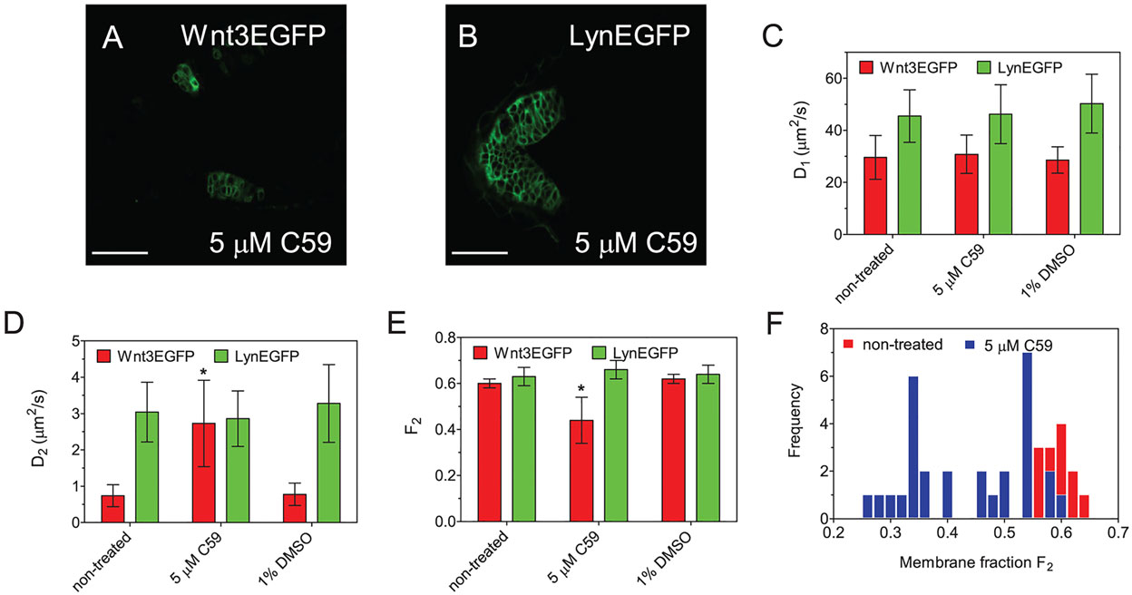
Fig. 7
Wnt3EGFP secretion is affected by the block of Porcupine. (A,B) Confocal images of zebrafish cerebellum expressing Wnt3EGFP and LynEGFP after C59 treatment. The embryos were treated with 5µM C59 10-28hpf (for details, see Materials and Methods). The samples were briefly soaked in 1× egg water before imaging in dorsal view, and FCS measurement. Scale bars: 50µm. (C-E) Diffusion coefficients (D1, D2) and protein membrane distribution (F2) extracted from fit at different conditions for both Wnt3EGFP and LynEGFP. (F) The results show that FCS signatures remain unchanged for LynEGFP, indicating that properties of the plasma membrane are not influenced by treatment or the drug function. Data are mean±s.d. Red bar, Wnt3EGFP; green bar, LynEGFP. Significance level, two-way t-test, *P<0.001. See also Table S3.