
Fig. S3
- ID
- ZDB-IMAGE-151207-7
- Publication
- Karra et al., 2015 - Myocardial NF-κB activation is essential for zebrafish heart regeneration
- All Figures
- Figures for Karra et al., 2015
|
Fig. S3
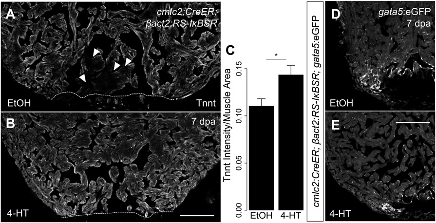
NF-KB signaling contributes to cardiomyocyte dedifferentiation. (A and B) Representative images of 7 dpa ventricles from cmlc2:CreER; βact2:RS-IKBSR treated with vehicle (n = 6) or 4-HT (n = 7) stained for Tnnt (gray). Arrowheads show diminished Tnnt staining in cardiomyocytes adjacent to the wound in control animals, suggestive of sarcomere disassembly. (C) Quantification of intensity of Tnnt intensity per muscle area suggests decreased sarcomere disassembly in ventricles with defective NF-KB signaling (n = 13). (D and E) Section images of 7 dpa ventricles from cmlc2:CreER; βact2:RS-IKBSR; gata5:eGFP fish treated with vehicle (n = 5) or 4-HT (n = 5). eGFP fluorescence is shown in grayscale. *P < 0.05, Student’s t test, two-tailed. (Scale bars: 100 µm.)