
Image
|
Figure Caption
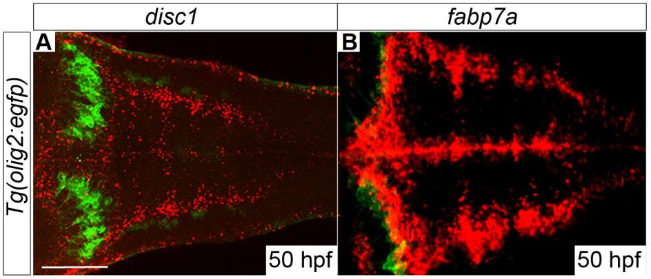
Fig. S1 Expression of disc1 in dorsolateral regions of the hindbrain resembles fabp7a/blbp expression. Fluorescent in situ hybridisation for disc1 (A, red) and fabp7a/blbp (B, red) was performed on Tg(olig2:EGFP) embryos in conjunction with immunofluorescent staining for EGFP (green) which identifies a population of cerebellar neurones seen as two prominent arcs at superficial levels of the hindbrain. The expression patterns of disc1 and fabp7a appear very similar in dorsolateral regions of the hindbrain.
Acknowledgments
This image is the copyrighted work of the attributed author or publisher, and
ZFIN has permission only to display this image to its users.
Additional permissions should be obtained from the applicable author or publisher of the image.
Full text @ Biol. Open