
Image
|
Figure Caption
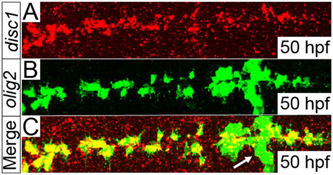
Fig. 4
disc1 is expressed in olig2-positive cells located around the midline of the hindbrain at 50hpf. A fluorescent in situ stain for disc1 mRNA (A, red) was performed in tandem with immunofluorescence staining for EGFP (B, green) on fixed Tg(olig2:egfp) embryos. The merged image shows clear overlap of the red and green signals in cells distributed along both sides of the midline of the hindbrain, but not in the abducens motor neurons (arrow). Dorsal views, anterior to left.
Figure Data
Acknowledgments
This image is the copyrighted work of the attributed author or publisher, and
ZFIN has permission only to display this image to its users.
Additional permissions should be obtained from the applicable author or publisher of the image.
Full text @ Biol. Open