
|
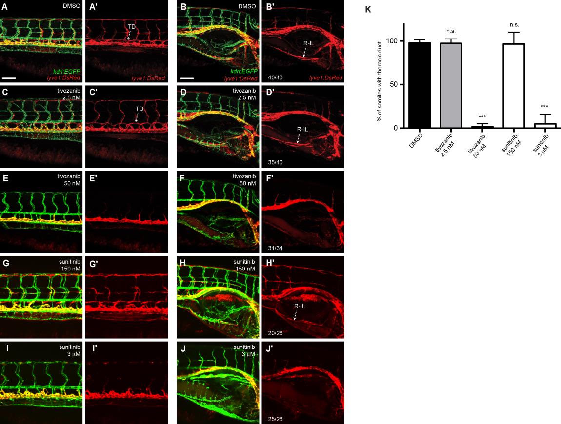
Fig. S4
Addition of 50 nM tivozanib or 3 uM sunitinib prevents the development of trunk and intestinal lymphatics. (A-J) Lateral images showing of 7 dpf lyve1:DsRed2;kdrl:EGFP embryos showing the trunk (A,C,E,G,I) and right intestinal (B,D,F,H,J) vasculature after treatment with 1% DMSO (A,B), 2.5 nM tivozanib (C,D), 50 nM tivozanib (E,F) 150 nM sunitinib (G,H) or 3 uM sunitinib (I,J) at 3 dpf (anterior to the right). (A′-J′) shows expression in the DsRed channel only. (K) Quantification of thoracic duct development in 7 dpf larvae treated with Vegfr inhibitors (ne25). Error bars, ±s.d. n.s P>0.05, ***P<0.001 by one-way ANOVA with Dunnett’s multiple comparison test. R-IL, right intestinal lymphatics; TD, thoracic duct. Scale bars: 100 µm.