
Image
|
Figure Caption
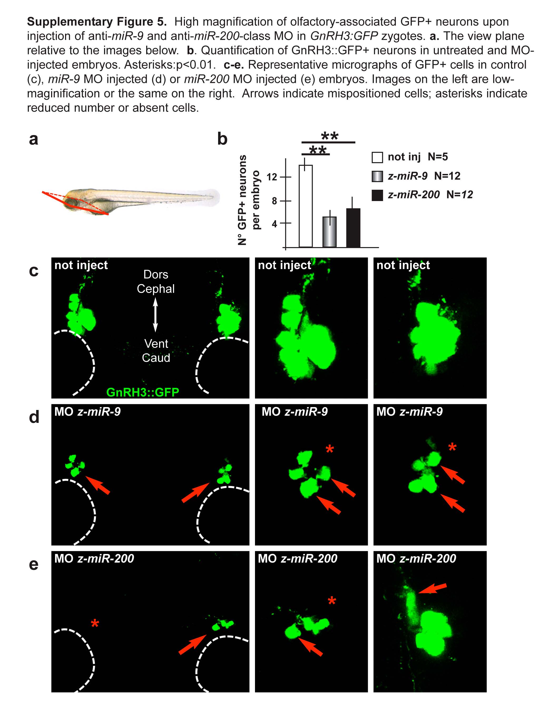
Fig. S5 High magnification of olfactory-associated GFP+ neurons upon injection of anti-miR-9 and anti-miR-200-class MO in GnRH3:GFP zygotes. a. The view plane relative to the images below. b. Quantification of GnRH3::GFP+ neurons in untreated and MO-injected embryos. Asterisks:p<0.01. c-e. Representative micrographs of GFP+ cells in control (c), miR-9 MO injected (d) or miR-200 MO injected (e) embyros. Images on the left are low-magnification or the same on the right. Arrows indicate mispositioned cells; asterisks indicate reduced number or absent cells.
Acknowledgments
This image is the copyrighted work of the attributed author or publisher, and
ZFIN has permission only to display this image to its users.
Additional permissions should be obtained from the applicable author or publisher of the image.
Reprinted from Molecular and cellular neurosciences, 68, Garaffo, G., Conte, D., Provero, P., Tomaiuolo, D., Luo, Z., Pinciroli, P., Peano, C., D'Atri, I., Gitton, Y., Etzion, T., Gothilf, Y., Gays, D., Santoro, M.M., Merlo, G.R., The Dlx5 and Foxg1 transcription factors, linked via miRNA-9 and -200, are required for the development of the olfactory and GnRH system, 103-19, Copyright (2015) with permission from Elsevier. Full text @ Mol. Cell Neurosci.