
|
Fig. 4
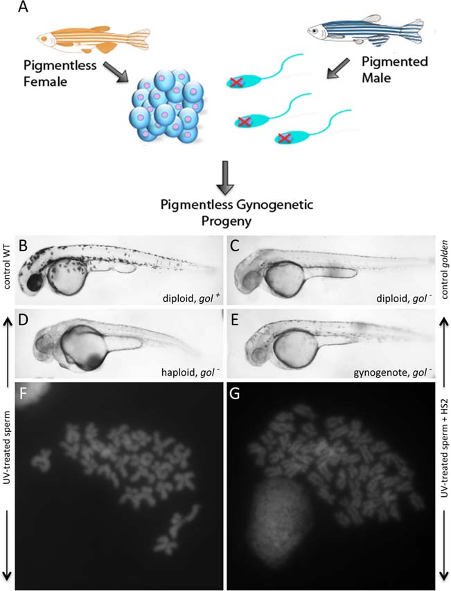
HS2 coupled to haploid production induces gynogenetic diploids. A: Diagram of the gynogenetic procedure. Maternal homozygosity for pigment markers is used to confirm that the resulting progeny lack genetic material from (wild-type pigmented) males. B–E: Phenotypes of 36 hpf embryos: (B) control wild-type (WT): diploid with normal (gol+) pigmentation, (C) control golden: wild-type diploid, homozygous for a mutation in the pigmentation gene golden (gol), which results in reduced levels of pigment in both melanophores and the eye, (D) haploid: haploid embryo derived from the fertilization of eggs from golden homozygous females with UV-treated sperm from wild-type males. The shortened body axis reflects the characteristic haploid phenotype. E: Gynogenote: gynogenetic diploid produced by treating haploid embryos (which would otherwise develop into embryos shown in C) with HS2 and selecting for a one-cycle division lag during the second cell cycle. In both D and E, golden pigmentation of the embryo indicates only maternal genetic material was transferred to the embryo. F,G: Chromosome counts in cells from 24 hpf embryos. F: Haploid chromosomal content in haploid embryos depicted in D. G: Diploid chromosomal content in gynogenetic diploids depicted in E.