
Image
|
Figure Caption
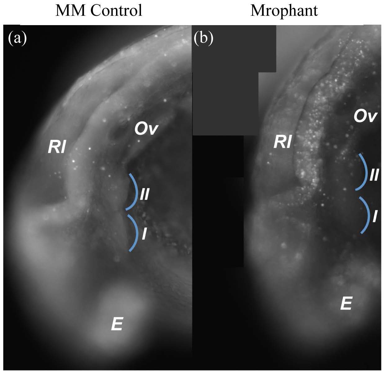
Fig. S4 Wnt9b morphants do not display increased apoptosis in the first pharyngeal arch. (a) (b) Lateral Views. (a) MM control and (b) morphant embryos were aged to 26hpf and subjected to acridine orange staining to assay cell death. Blue arcs indicate PA1&2. Note that apoptotic cells within the PAs are not increased in morphants. Apoptotic cells within dorsal neural structures are increased in morphants. Br = brain, E = eye, Ov = otic vesicle, Rl = rhombic lips.
Acknowledgments
This image is the copyrighted work of the attributed author or publisher, and
ZFIN has permission only to display this image to its users.
Additional permissions should be obtained from the applicable author or publisher of the image.
Full text @ Am. J. of Molec. Bio.