
Image
|
Figure Caption
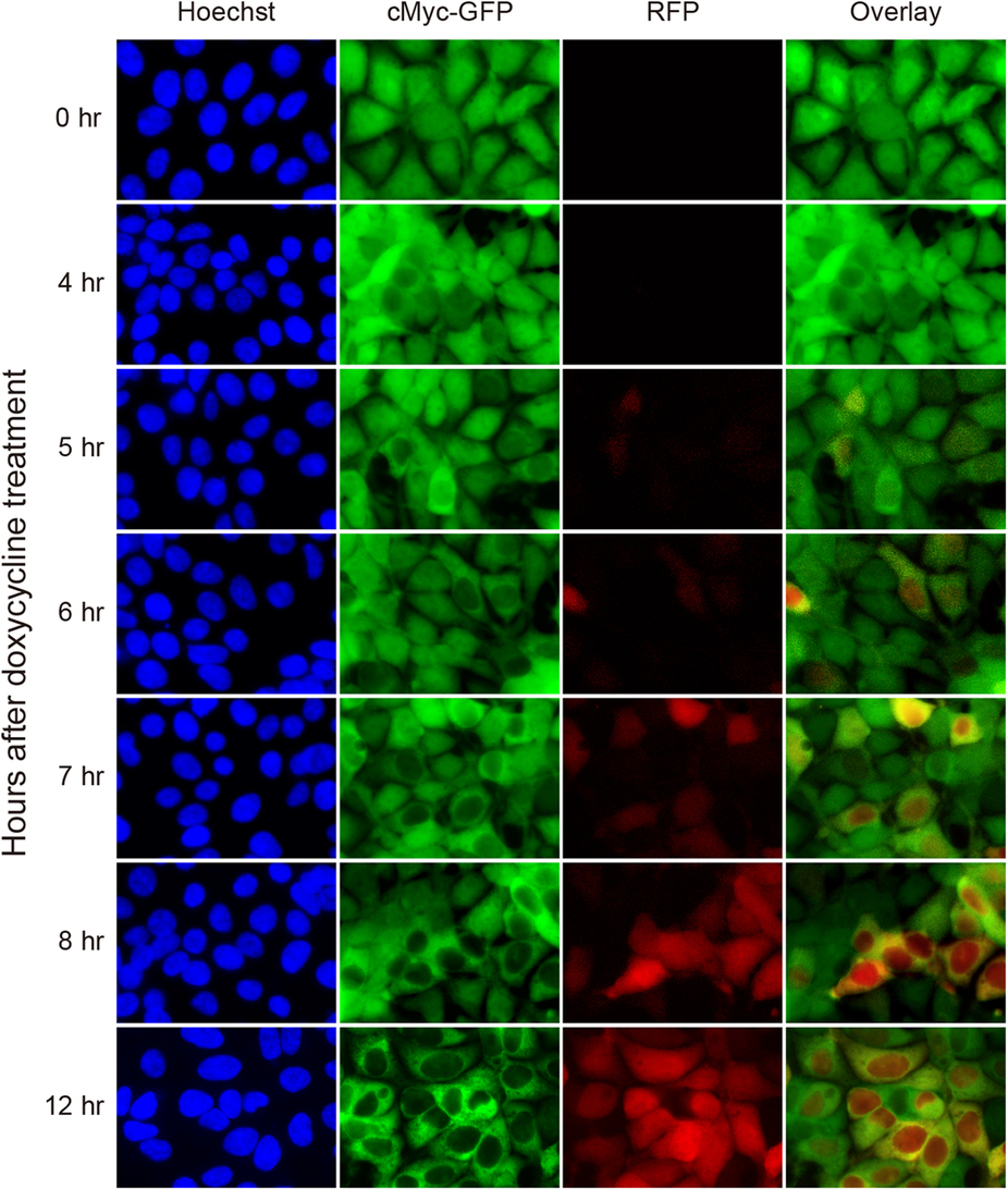
Fig. 4
Time course of nuclear cMyc-GFP depletion by Ab-SPOP.
293TetOn cells stably expressing cMyc-GFP from a CMV promoter and RFP/Ab-SPOP from a bi-directional TRE promoter were imaged under epifluorescence at different time points (0, 4, 5, 6, 7, 8, 12 h) after doxycycline (1 µg/ml) administration. Overlay merges images of c-Myc-GFP and RFP.
Acknowledgments
This image is the copyrighted work of the attributed author or publisher, and
ZFIN has permission only to display this image to its users.
Additional permissions should be obtained from the applicable author or publisher of the image.
Full text @ Sci. Rep.