
Image
|
Figure Caption
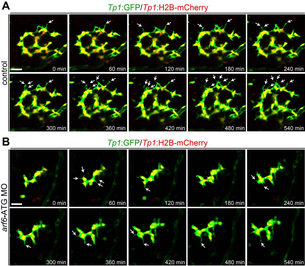
Fig. S6
The movement of BEC filopodia is greatly reduced in arf6-ATG MO-injected larvae.
(A, B). Time-lapse confocal images showing BEC behaviors in arf6-ATG MO-injected and control larvae. The behaviors were assessed by Tp1:GFP (green) and Tp1:H2B-mCherry (red) expression in BEC cytoplasm and nuclei, respectively. Confocal images every 60 minutes from 74 to 84 hpf were presented. Arrows point to BEC filopodia. Scale bars, 25 µm.
Figure Data
Acknowledgments
This image is the copyrighted work of the attributed author or publisher, and
ZFIN has permission only to display this image to its users.
Additional permissions should be obtained from the applicable author or publisher of the image.
Full text @ PLoS One