
|
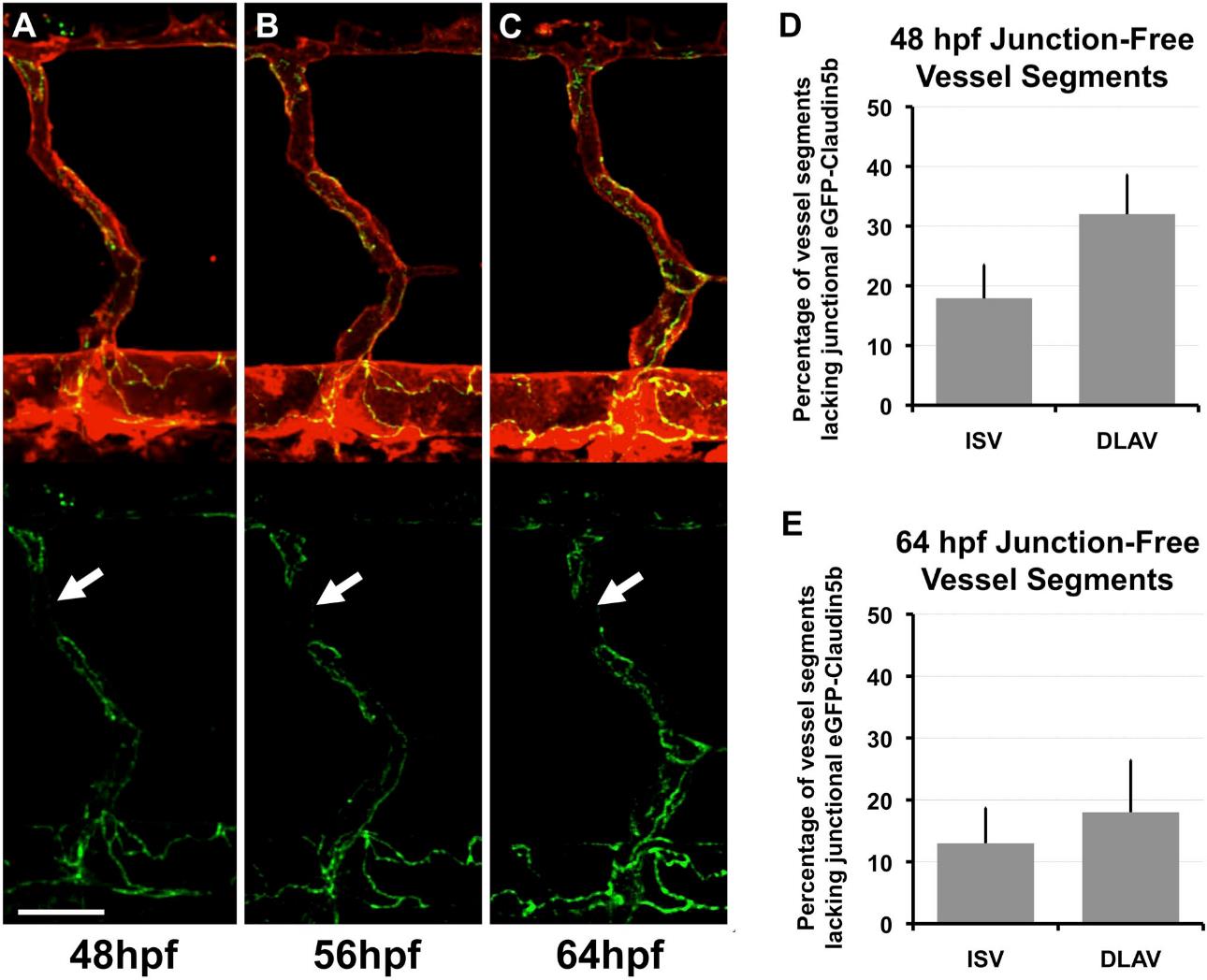
Fig. S5 (A-C) Confocal micrographs of mRFP/EGFP (top; EC membranes in red and junctions in green) and EGFP (bottom; junctions in green) fluorescence in the same vascular segments in a Tg(fli1a:egfp-claudin5b)y287; Tg(kdrl:mRFP-F)y286 double transgenic animal at 48 hpf (A), 56 hpf (B), and 64 hpf (C). Tight junction-free vessel segments (arrows) persist for at least a day after intersegmental vessel lumenization. (D,E) Quantification of the percentage of vessel segments lacking junctional EGFP-Claudin5b, measured in the same Tg(fli1a:egfp-claudin5b)y287; Tg(kdrl:mRFP-F)y286 double transgenic animals at 48hpf (D) and 64 hpf (E). Although junction-free segments persist, the overall length of these segments does decrease over time. Scale bar = 20 µm.