
|
Fig. S1
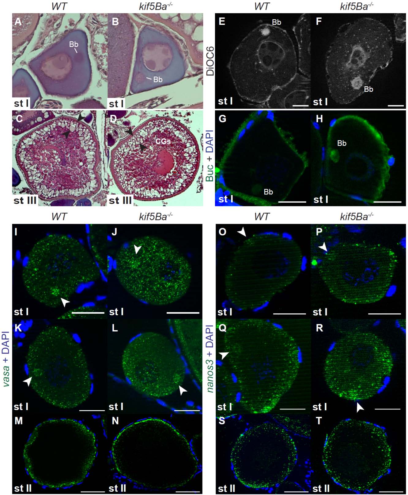
kif5Ba is dispensable for oocyte polarity and germ plasm localization.
(A-D) H&E staining of WT (A,C) and kif5Ba-/- mutant (B,D) oocytes showing polarized stage I (st I) oocytes with Balbiani bodies (Bb) and normal stage III (st III) oocytes with cortically-localized cortical granules (CGs, between arrowheads) and centrally localized nucleus and yolk. (n=3 mutant ovaries analyzed, one each of kif5Baae11/ae11, kif5Baae11/ae12, and kif5Baae12/ae12; assayed 5-10 oocytes for each genotype).
(E-F) DiOC6 staining reveals that ER and mitochondria are properly localized to the Bb in WT (E) and kif5Ba-/- mutant (F) st I oocytes. (n=18 oocytes analyzed from kif5Baae11/ae11 mutant ovary, n=19 oocytes analyzed from WT ovary). Scale bars 20µm.
(G-H) Buc is properly localized to the Bb in WT (G) and kif5Ba-/- mutant (H) st I oocytes. (n=12 oocytes analyzed from kif5Baae11/ae11 mutant ovary, n=10 oocytes analyzed from WT ovary)). Scale bars 20µm.
(I-T) vasa (I-N) and nanos3 (O-T) RNA are enriched in the Bb (arrowheads) in early st I (I,J,O,P) and late st I (K,L,Q,R) oocytes in WT (I,K,O,Q) and kif5Ba-/- mutants (J,L,P,R). vasa RNA localizes to the cortex in WT (M) and kif5Ba-/- mutant (N) st II oocytes. nanos3 RNA is diffuse throughout the cytosol of WT (S) and kif5Ba-/- mutant (T) st II oocytes. (n=3 WT and kif5Baae12/ae12 mutant ovaries, assayed ≥10 oocytes for each condition). Scale bars 25µm (I-L, O-R), 25µm (M,N,S,T).