
|
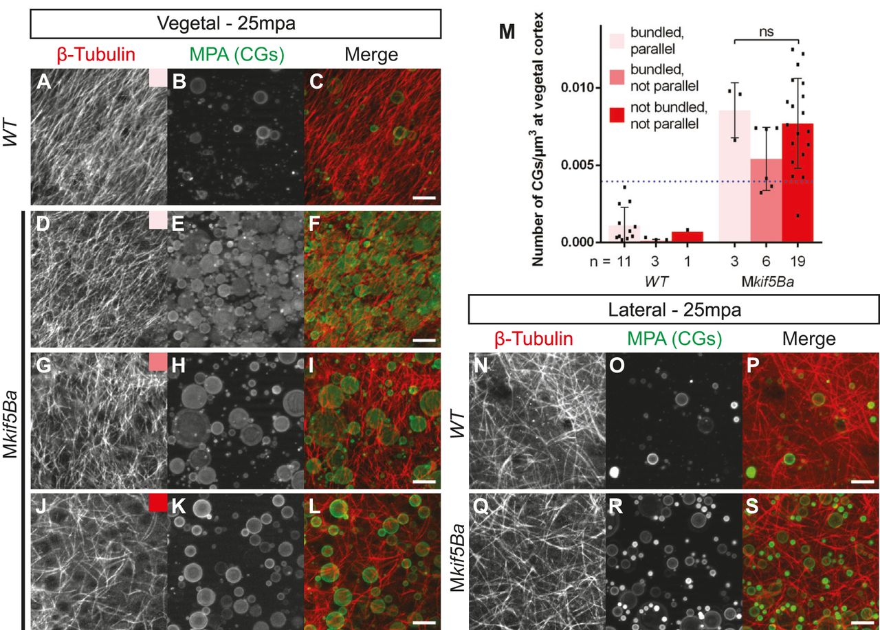
Fig. 7
Maternal kif5Ba organizes vegetal microtubules. (A-M) Vegetal microtubules are in a parallel orientation and bundled in WT activated eggs (A), but in Mkif5Ba mutants are either bundled and parallel (D), bundled but not parallel (G), or not bundled or parallel (J). Mkif5Ba mutant activated eggs display variable CG exocytosis defects (compare B with E,H,K). Importantly, the degree of CG retention does not correlate with the severity of the microtubule phenotype in Mkif5Ba mutants (M). Scale bars: 10µm. Blue dotted line in M represents the upper limit of CG retention observed in WT activated eggs. Error bars show mean±s.d.; one-way ANOVA. (N-S) There is no difference between lateral microtubule organization in WT (N-P) and Mkif5Ba mutant (Q-S) activated eggs. Scale bars: 10µm.