
|
Fig. 1
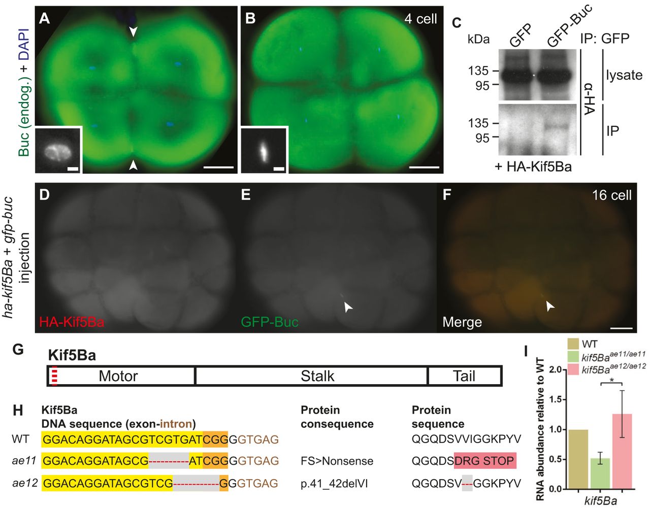
Endogenous Buc localizes to the germ plasm of embryos and binds Kinesin-1. (A,B) Endogenous Buc localizes to distal cleavage furrows of 4-cell embryos when chromosomes are decondensed (A), but not during metaphase (B) or anaphase. Insets show DAPI. Arrowheads indicate Buc accumulation. Scale bars: 100µm for main images, 10µm for insets. See text for quantification. (C) GFP-Buc, but not GFP, co-immunoprecipitates HA-Kif5Ba in HEK293T cells. (D-F) Overexpressed GFP-Buc localizes to cleavage furrows and overexpressed HA-Kif5Ba is expressed throughout blastomeres, potentially allowing them to interact in vivo. Arrowheads indicate Buc accumulation. (G) Schematic protein structure of Kif5Ba, illustrating motor, stalk and tail domains. Red dashed line indicates CRISPR target site. (H) DNA and predicted protein sequences of WT and mutant kif5Ba alleles (ae11 and ae12). Yellow indicates protospacer, orange indicates PAM, gray area/red dashed line indicates a deletion and pink indicates altered amino acids. (I) qRT-PCR for kif5Ba from pools of 5-dpf WT and homozygous mutants. kif5Ba undergoes nonsense-mediated decay in kif5Baae11/ae11 but not kif5Baae12/ae12 mutants. Error bars show mean±s.e.m.; Student′s t-test, *P=0.0388.