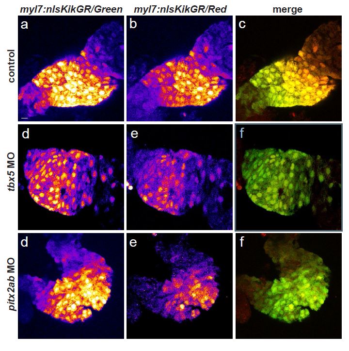
Fig. S6 Impact of tbx5a and pitx2ab knockdown on SHF addition to the FHF. Scale bar = 10 µm. (a-i) Photoconversion-mediated lineage tracing of the SHF cardiomyocytes addition to the linear heart tube. Photoconversion of cardiomyocyte-specific Tg(myl7:nlsKikGR) was performed at 365 nm for 2 minutes at 27 hpf and the hearts imaged at 56 hpf; the total conversion to RFP was visually confirmed. a, d, g, shows the green channel, b, e, h, shows the red channel, merge is shown in c, f, i. All GFP-positive nuclei are the newly added cardiomyocytes after 27hpf, RFP-labeled nuclei represents all cardiomyocytes of the linear heart tube prior to photoconversion, yellow in merge (c, f, i). a-c, control hearts, d-f, hearts from tbx5a morphants show mild decrease in newly added cardiomyocytes, g-i, hearts from pitx2ab morphants display increase in cardiomyocyte recruitment.
                
                    
                        Image


                        Figure Caption


                        Acknowledgments


This image is the copyrighted work of the attributed author or publisher, and
ZFIN has permission only to display this image to its users.
Additional permissions should be obtained from the applicable author or publisher of the image.
      Full text @ Nat. Commun.