
|
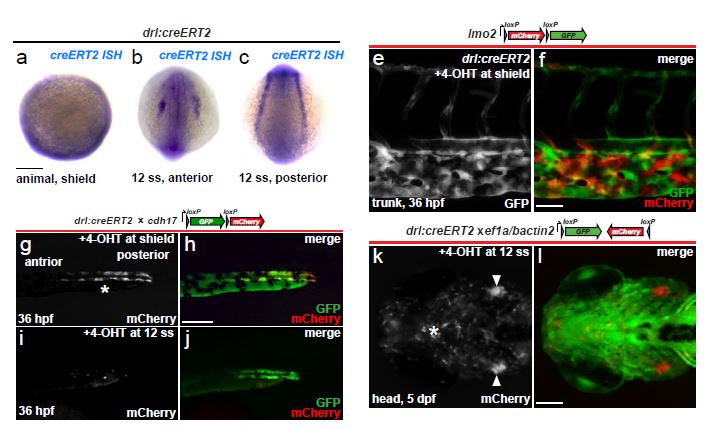
Fig. S2 drl reporter expression refines to anterior and posterior cardiovascular lineages over time. (a-c) mRNA in situ hybridization (ISH) for creERT2 in drl:creERT2 embryos shows expression at shield stage (a, animal pole view, ventral on top) at the embryo margin, and at 12 somite stage (b, c) shows the separation of drl reporter expression into anterior and posterior lateral mesoderm; scale bar = 100 µm (e, f) Vascular lineage tracing confirmed using drl:creERT2 crossed to the endothelial-specific loxP reporter lmo2:Switch (schematic) and 4- OHT-induced at shield stage, tracing (GFP signal, green) shown in the trunk at 36 hpf; scale bar = 50 µm. (g-j) Transgenic zebrafish carrying drl:creERT2 crossed to cdh17:loxP-GFP-loxP-mCherry (schematic) were induced with 4-OHT at shield or 12 somite stage; mCherry expression in the renal tubules reveals drl expression in their precursors during gastrulation (g, h), but not at later stages of somitogenesis (i, j); scale bar = 100 µm. (k, l) Definitive hematopoietic lineages descend from drl-expressing cells at 12 ss, as shown in a 5-day larva with tracing to the developing bilateral thymus (arrowheads, k) and tissue-infiltrating leukocytes (asterisks, k) using the ef1a/beta-actin2-based FlEx reporter line (schematic); scale bar = 100 µm.