
|
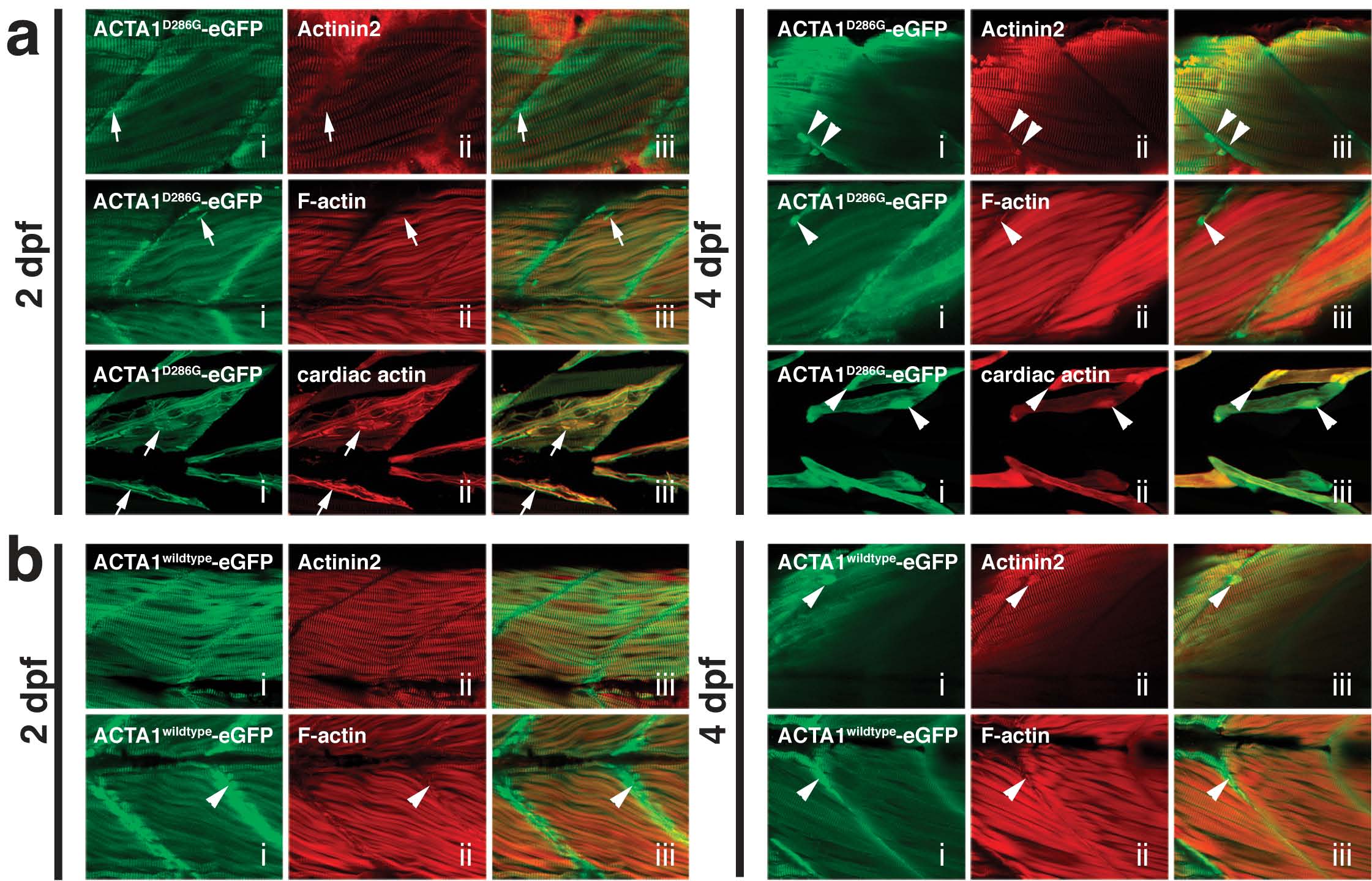
Fig. S2 Characterization of nemaline bodies and aggregates in Tg(ACTA1-eGFP)high zebrafish. a) Confocal microscopy images of nemaline bodies and aggregates in Tg(ACTA1D286G-eGFP)high muscle. At 2 dpf, nemaline bodies (arrows) in (i) Tg(ACTA1D286G-eGFP)high muscle (green) do not stain with Actinin2 (red; ii and overlaid in iii) or phalloidin (labeling F-actin) (red; i and overlaid in ii) despite correct localization of these markers in the sarcomere. At 4 dpf, aggregates (arrowheads) in (i) Tg(ACTA1D286G-eGFP)high muscle (green) stain for Actinin2 (red; ii and overlaid in iii), and phalloidin (red; ii and overlaid in iii) in Tg(ACTA1D286G-eGFP)high muscle (green). Co-injection of actc1b;ACTA1D286G-eGFP and actc1b;actc1a-mCherry (cardiac α-actin) results in mosaic expression of both constructs throughout the skeletal muscle. ACTA1D286G-eGFP (green) and cardiac actin (red) co-localize in nemaline bodies at 2 dpf (arrows; ii and overlaid in iii) and in aggregates at 4 dpf in (arrowheads; ii and overlaid in iii). b) Confocal microscopy images of globular aggregates in Tg(ACTA1wildtype-eGFP)high muscle. At 2 dpf (arrows), and 4 dpf, aggregates (arrowheads) are observed in (i) Tg(ACTA1wildtype-eGFP)high skeletal muscle (green) labeled with Actinin2 (red; ii and overlaid in iii) and phalloidin (red; ii and overlaid in iii).