
|
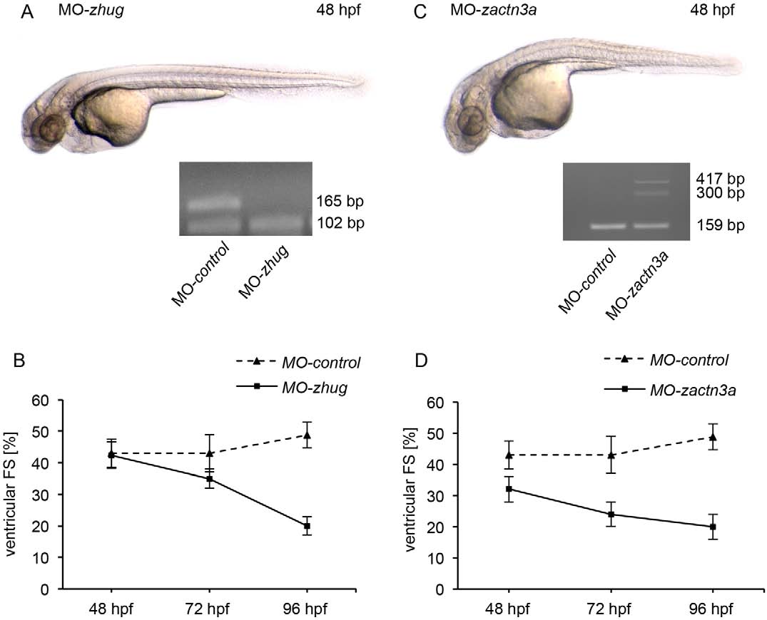
Fig. 6
Correctly balanced huG and actn3a isoform expression is essential for proper heart function. (A,B) Lateral view of a MO-huG-injected (MO-zhug) zebrafish embryo at 48hpf. Fractional shortening (FS) shows a progressive reduction of cardiac contractility. Inset, cDNA splice-site analysis of huG morphant. (C,D) Splice-site blockage in actn3a leads to mild cardiac dysfunction. actn3a-depleted zebrafish embryos develop heart failure with dilation of the atrium and reduced ventricular contractility beginning at the 72h developmental stage. Inset, cDNA splice-site analysis of actn3a morphant. Results in B and D are mean±s.d. (n=10).