
|
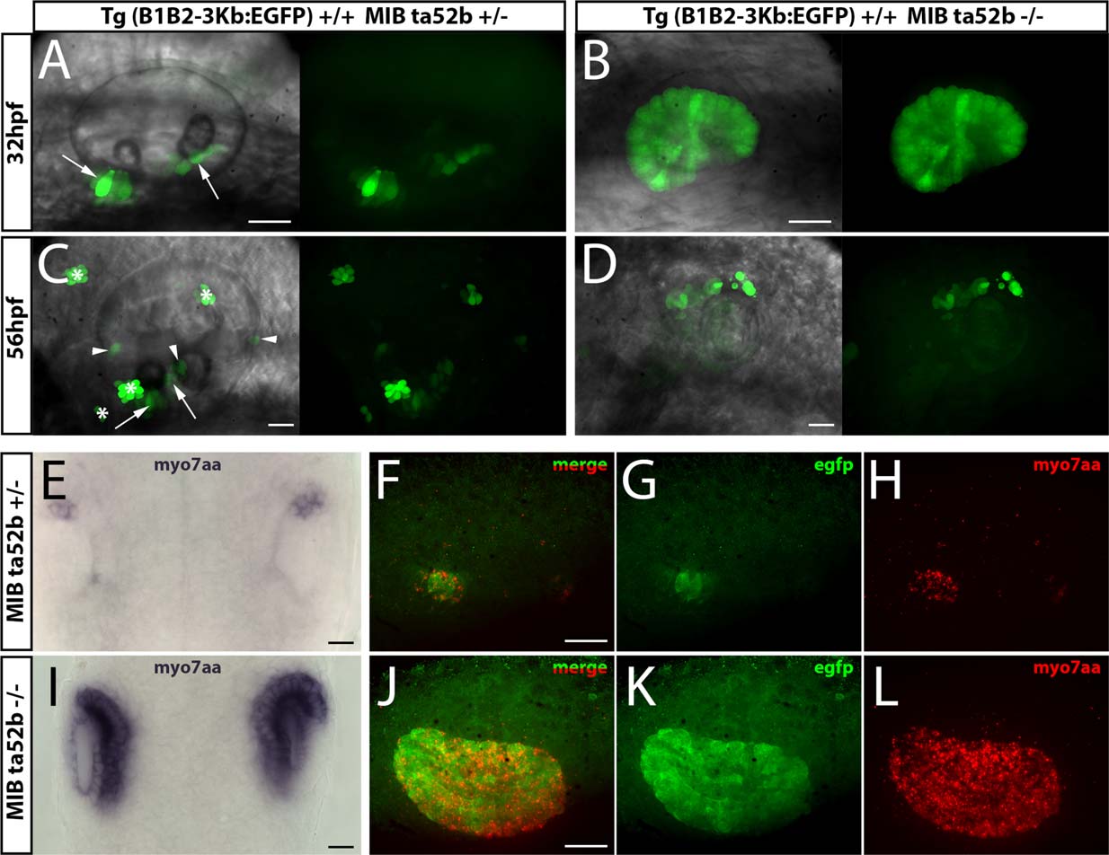
Fig. 6
Element B1B2-3Kb responds to MIB disruption by an enlargement of EGFP expression area. Expression of EGFP in fish transgenic for the B1B2-3Kb DNA fragment and carrying heterozygous (A,C) or homozygous (B,D) MIB ta52b mutation. Live embryos were anesthetized with tricaine and mounted in agarose before imaging on a Leica spinning disk microscope. All images are z-axis projections. Embryonic stages imaged were: 32 hpf (A,B), 56 hpf (C,D). All panels comprise the superimposition of bright field with fluorescence image (left part) and the fluorescence image (right part) of the exact same area of the embryo. A,C: arrows = anterior and posterior maculae; arrowhead = anterior, lateral and posterior cristae; * = neuromast. Enlargement of EGFP expression domain in MIB ta52b mutants carrying B1B2-3Kb:EGFP transgene correlates with that of myo7aa mRNA. Detection of endogenous myo7aa mRNA in Tg(B1B2-3Kb:EGFP)+/+ MIB ta52b +/ (E-H) and Tg(B1B2-3Kb:EGFP)+/+ MIB ta52b / (I–L) embryos at 24 hpf. (E,I) dorsal view of embryos showing in situ hybridization staining of myo7aa mRNA revealed with NBT/BCIP in the two otic vesicles. (F–H, J–L) double staining for EGFP (immunofluorescence) and myo7aa mRNA (FastRed in situ hybridization) in MIB ta52b +/ (F–H) and MIB ta52b / carrying B1B2-3Kb:EGFP transgene (J–L). Merged fluorescence (F,J), EGFP (G,K) and myo7aa (H,L) visualized in lateral views of otic vesicle. Scale bar: 20 µm (A–L).