
Image
|
Figure Caption
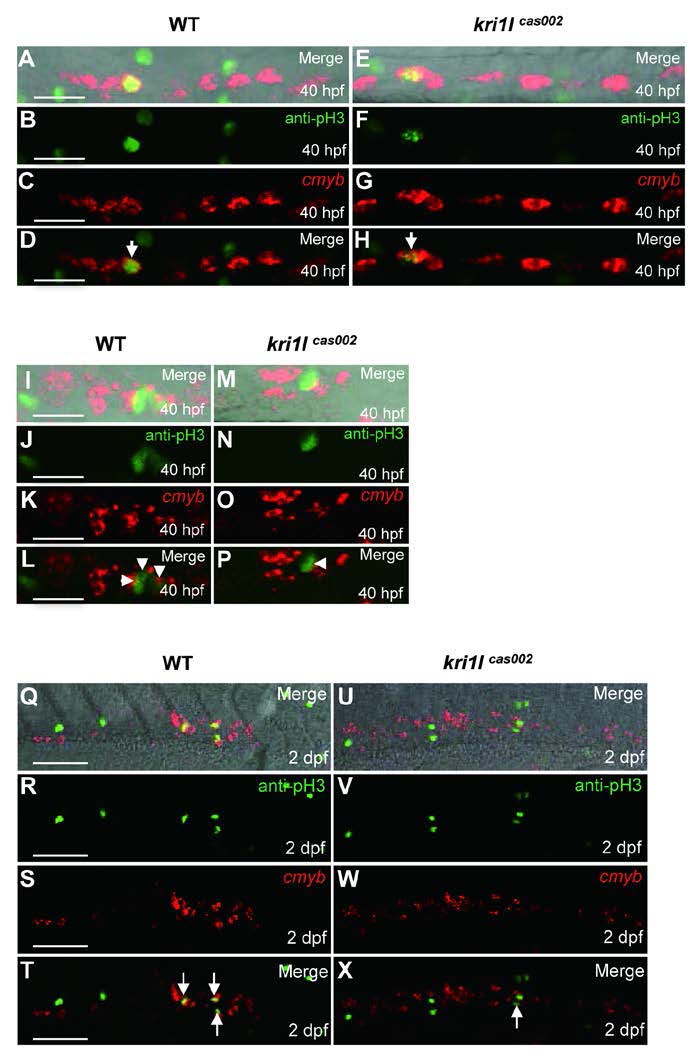
Fig. S10 Defective HSPC proliferation in kri1lcas002 mutants. Representative confocal images of double staining of cmyb WISH (red) and phospho-Histene 3 immunostaining (green) in different time points and regions, including the AGM at 40 hpf (A-H) (A and E are bright-field images overlaid with fluorescent staining. Scale bars, 25 µm.); the CHT at 40 hpf (I-P) (I and M are bright-field images overlaid with fluorescent staining. Scale bars, 25 µm.), and at 2 dpf (Q-X) (Q and U are bright-field images overlaid with fluorescent staining. Scale bars, 50 µm.).
Acknowledgments
This image is the copyrighted work of the attributed author or publisher, and
ZFIN has permission only to display this image to its users.
Additional permissions should be obtained from the applicable author or publisher of the image.
Full text @ Cell Res.