
|
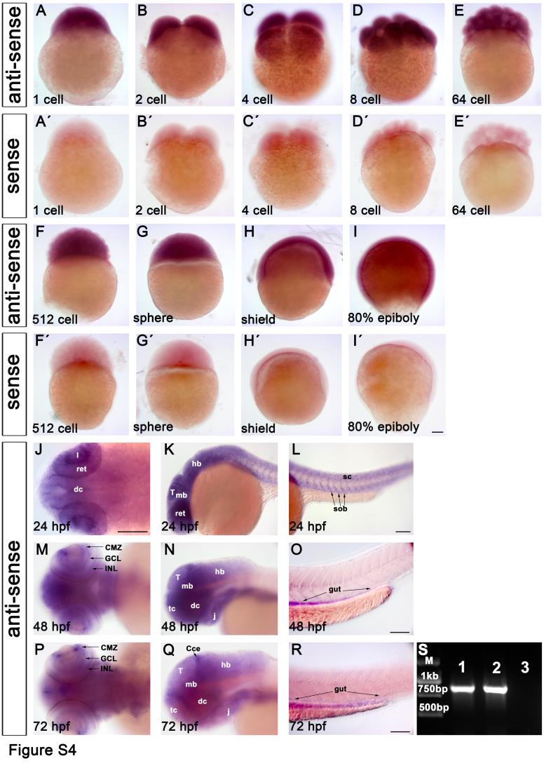
Fig. S4
Characterization of ddb1 expression pattern during wild type zebrafish embryonic development by WISH and RT-PCR.
(A-I′) The WISH signal of ddb1 antisense probe (A-I) and its sense control (A′-I′) in embryos before 24 hpf. ddb1 transcript was ubiquitously detected in all blastomeres (A-F′) before mid-blastula transition (MBT) when zygotic transcription starts, revealing that ddb1 was expressed maternally. The ubiquitous expression was continued in subsequent stages including sphere, shield and 80% epiboly (G-I, G′-I′). (J-R) The expression of ddb1 at 24 hpf (J-L), 48 hpf (M-O), and 72 hpf (P-R). From 24 hpf onwards, ddb1 mRNA was observed to be expressed broadly and at high levels in the brain and somites (J-L), then spatially restricted to the brain, retina (high in the CMZ but moderate in the GCL and INL, M), the branchial arches, and endoderm at 48 hpf (N-O), followed at 72 hpf by downregulation and more distinct spatial expression pattern (P-R). ddb1 was detected at moderate levels in the telencephalic proliferation region, tectal proliferation region, cerebellum, CMZ, and branchial arches, whereas in other regions the signal was weak (P-Q). (S) ddb1 was maternally expressed as revealed by RT-PCR from two separate cDNA templates prepared from one cell stage zygotes (lanes 1 and 2; lane 3—control with water only as template) using a ddb1 specific primer pair.