
Image
|
Figure Caption
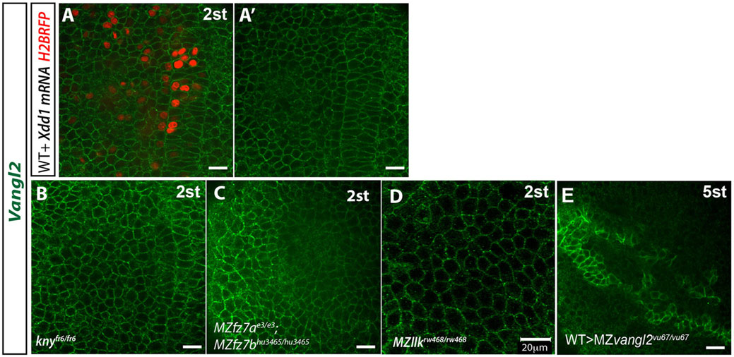
Fig. 5
Vangl2 membrane localization in PCP mutant backgrounds. Confocal images of whole-mount immunostaining with antibody against zebrafish Vangl2 C-terminus at 2s stage (10.7hpf) in embryos mosaically expressing Xdd1 and H2B-RFP (A,A′) or in knyfr6/fr6 (B), MZfz7ae3/e3;MZfz7bhu3465/hu3465 (C), MZllkrw468/rw468 (D) or MZvangl2vu67/vu67 embryos (11.7hpf, 5 s) containing transplanted WT cells (showing strong Vangl2 membrane expression) (E). Scale bars: 20µm.
Acknowledgments
This image is the copyrighted work of the attributed author or publisher, and
ZFIN has permission only to display this image to its users.
Additional permissions should be obtained from the applicable author or publisher of the image.
Full text @ Development