
Fig. 7
- ID
- ZDB-IMAGE-150929-16
- Publication
- Tsai et al., 2015 - Modulation of p53 and met expression by krüppel-like factor 8 regulates zebrafish cerebellar development
- All Figures
- Figures for Tsai et al., 2015
|
Fig. 7
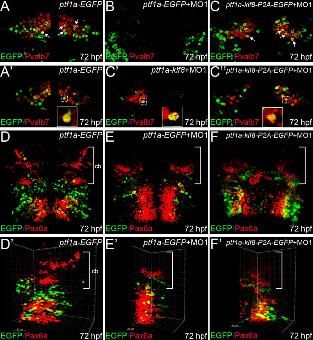
Klf8 controls formation of cerebellar Purkinje cells in a cell-autonomous manner. (A–C′′) Examination of wild type embryos injected with ptf1a-EGFP at 72 hpf revealed that differentiated Purkinje cells express Pvalb7 (red), and that some Purkinje cells (derived from ptf1a:EGFP cells) express both EGFP and Pvalb7 (yellow, arrows) (A, A′). Examination of embryos co-injected with klf8-MO1atg and ptf1a-EGFP revealed that no Purkinje cells had formed by 72 hpf (B). In embryos co-injected with klf8-MO1atg and ptf1a-klf8-P2A-EGFP, some ptf1a:klf8-P2A-EGFP+ cells differentiated into Purkinje cells expressing Pvalb7 and EGFP (yellow, arrows), while ptf1a promoter activity was switched off in the majority of Pvalb7+ Purkinje cells (red) derived from ptf1a: klf8-P2A-EGFP+ cells, so that these cells no longer expressed EGFP at 72 hpf (C; C′, n = 5; C′′, n = 5). (D–F′) In wild type embryos injected with ptf1a-EGFP, granule cells expressing Pax6a (red, bracket) are observed in the cerebella of embryos at 72 hpf (D, D′). Examination of embryos co-injected with klf8-MO1atg and ptf1a-EGFP revealed reduced numbers of granule cells expressing Pax6a (red) in the cerebella of 72 hpf embryos (E, E′). Examination of embryos co-injected with klf8-MO1atg and ptf1a-klf8-P2A-EGFP revealed a similar reduction in the number of granule cells expressing Pax6a (red) in the cerebella of embryos at 72 hpf (F, F′). Dorsal views of embryos are shown. Z-axis merged images are shown in panels A–C and D–F′. Single confocal images are shown in panels A′, C′, and C′′. Panels D′–F′ are the same images as shown in D–F, but rotated clockwise by ~30–45°. Cb, cerebellum.