
|
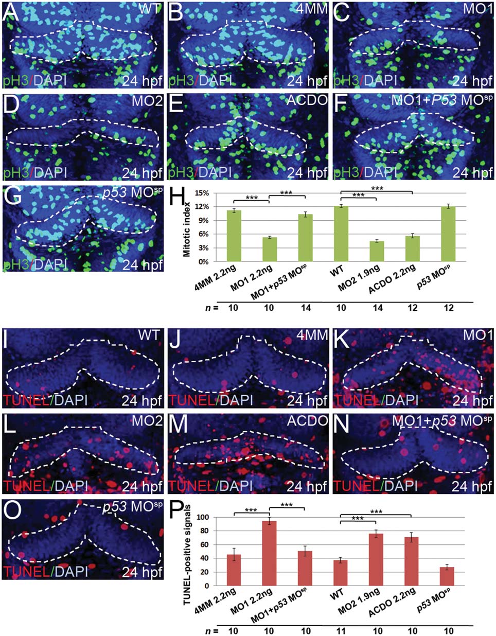
Fig. 3
Knockdown of klf8 reduces cell proliferation and increases apoptosis in the cerebellar anlage. (A–G) Dorsal views of embryos. The number of pH3-expressing M phase cells in the cerebellar primordia at 24 hpf is decreased in embryos injected with klf8-MO1atg (C), klf8-MO2atg (D), or co-injected with klf8-MOAC and klf8-MODO (E), as compared with WT (A) or embryos injected with klf8-4mmMO1 (B) or p53 MOsp (G). Co-injection of p53 MOsp with klf8-MO1atg rescues the cell proliferation defect (F). (H) Mitotic indices of embryos of the indicated backgrounds. (I–O) Dorsal views of embryos. Increased apoptosis in the cerebellar primordium at 24 hpf is observed in embryos injected with klf8-MO1atg (K), klf8-MO2atg (L), or co-injected with klf8-MOAC and klf8-MODO (M), as compared with WT (I), or embryos injected with klf8-4mmMO1 (J) or p53 MOsp (O). Co-injection of p53 MOsp with klf8-MO1atg rescues the apoptosis defect (N). (P) Percentage of TUNEL-positive cells in embryos of the indicated backgrounds. Significance was determined using Student′s t-test. ***p < 0.001. Error bars indicate standard errors.