
|
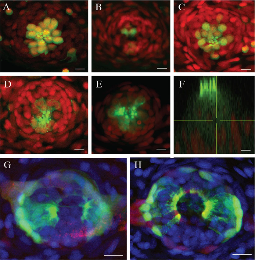
Fig. 2
Efficient hair-cell regeneration in adult zebrafish. (A-C) Maximal projection of confocal images from Tg[ET(krt4:EGFP)sqet4] transgenics (green) showing neuromasts of the caudal fin of a 2-year old fish stained with DAPI (red) (A) before neomycin-treatment, (B) 2h after treatment, and (C) 72h after treatment. (D-E) Neuromast of an adult fish from Tg[myo6b:actb1-EGFP] transgenics (green) stained with DAPI (red) showing (D) hair cells controls, and (E) in neomycin-treated fish after hair-cell regeneration. (F) XZ profile of the same neuromast in E showing the apicobasal polarization of the regenerated hair cells (green). (G-H) Neuromast of an adult fish from Tg[Alpl:mCherry (red) ; ET(krt4:EGFP)sqet20 (green)] transgenics stained with DAPI (blue), showing (G) epithelial geometry in control fish and (H) in neomycin-treated fish at 72hpt. In all cases, N=8 neuromasts from 2 animals. Scale bars=10µm.